Verret, L. et al. (2012) Cell 149, 708.
In this featured paper, we discuss the work of Verret, L. et al. which aims at understanding the mechanisms leading to the cognitive decline and network change in Alzheimer’s disease (AD). Western blot analysis of brain lysates from human AD subjects and mouse AD model system using Alomone Labs Anti-SCN1A (NaV1.1) Antibody (#ASC-001), Anti-SCN2A (NaV1.2) Antibody (#ASC-002), Anti-SCN3A (NaV1.3) Antibody (#ASC-004) and Anti-NaV1.6 (SCN8A) Antibody (#ASC-009), showed that NaV1.1 channel levels are decreased. NaV1.1 may have a role in AD.
Alzheimer's disease (AD) is a slowly progressing neurological disease that reduces life expectancy and diminishes the quality of life. AD has both sporadic and autosomal-dominant occurrences that irreversibly impair cognition and lead to debilitating dementia that is sometimes accompanied by epileptic outbursts. Among the many contributors to AD pathogenesis is the increase in amyloid-β (Aβ) plaques in the proximity to active glutamatergic synapses, are perceived as a determining factor in the onset of the disease. Indeed, transgenic mice with enhanced Aβ expression (human amyloid precursor protein, or hAPP, lines) show AD related symptoms such as behavioral aberrancies and spontaneous epileptiform seizures. Here, the hypothesis that hyper-synchronous network activity, observed as irregular gamma wave oscillations, (i.e. epileptic episodes), is linked to symptomatic AD has prompted Verret et al. to conduct this reviewed study.
Using electroencephalograph (EEG), the authors monitored parietal cortices of hAPP mice; they revealed reverse relationship between gamma oscillation intensity and epileptic spikes. Gamma oscillations are generated by synchronous activity of inhibitory parvalbumin (PV) cells and are required to maintain responsive neuronal circuitry; indeed, hAPP mice with enhanced PV functionality and/or gamma activity outperform hAPP mice with increased spike frequency in behavioral tests, whereas the antiepileptic drugs riluzole and phenytoin (which are specific VGSCs blockers) reduced gamma activity in hAPP mice and therefore aggravated epileptiform events. Moreover, spontaneous inhibitory post synaptic currents (sIPSCs), from layer II/III GABAergic synapses were significantly decreased in hAPP than in wild type (WT) mice, in the presence of action potentials (AP). Further examination of the intrinsic properties of PV cells confirmed that fast spiking GABAergic interneurons of hAPP mice were more depolarized and lower in action potential amplitudes, relative to WT mice.
Voltage-gated Na+ channels (VGSCs) are known to mediate AP initiation and progression and have distinct expression patterns across the CNS. Western blot analyses using Anti-SCN1A (NaV1.1) Antibody (#ASC-001), Anti-SCN2A (NaV1.2) Antibody (#ASC-002), Anti-SCN3A (NaV1.3) Antibody (#ASC-004), and Anti-NaV1.6 (SCN8A) Antibody (#ASC-009), corresponding to the most common Na+ channels in the CNS, show reduced levels of NaV1.1 and NaV1.6 (but not the other VGSCs subtypes mentioned) in the parietal cortex of both hAPP mice and AD patients, compared with WT and non-demented controls, respectively (Figure 1). Following this observation, in situ hybridization of the parietal cortex, showed that both NaV1.1 and NaV1.6 channels reside primarily on PV cells, but to a much lesser degree in hAPP mice than in WT mice.
Figure 1. NaV1.1 Levels Decrease in PV Cells of hAPP Mice and in AD Brains.A and B. Western blot analysis of parietal cortex from mice and inferior parietal cortex from humans using Anti-SCN1A (NaV1.1) Antibody (#ASC-001), Anti-SCN2A (NaV1.2) Antibody (#ASC-002), Anti-SCN3A (NaV1.3) Antibody (#ASC-004) and Anti-NaV1.6 (SCN8A) Antibody (#ASC-009) antibodies. The graphs represent the quantitation of each western blot.Adapted from Verret, L. et al. (2012) with permission of Elsevier.
To compensate for the VGSCs deficiency in PV cells of hAPP mice and thereby to better understand the role of these channels in asynchronous gamma waves, the authors crossed hAPP mice with mice overexpressing NaV1.1, thereby restoring or increasing NaV1.1 levels in PV cells. in situ hybridization of PV cells showed that hAPP/NaV1.1 mice had NaV1.1 levels similar to WT mice. Subsequent EEG recordings were consistent with the authors’ hypothesis: parietal cortices of hAPP/NaV1.1 mice had lengthened high-intensity gamma activity episodes, and lacked abnormalities in sIPSCs. Further, these mice had reduced epileptic discharges and baseline gamma activity.
Lastly, behavioral paradigms showed that hAPP/NaV1.1 mice ranked higher in habituation to a novel environment and had improved spatial learning and memory. Overall, this study highlights the role of VGSCs in AD, and emphasizes the importance of NaV1.1 in the pathogenesis of this disease.
Kim, S. et al. (2012) Nat. Neurosci. 15, 1236.
The olfactory system of vertebrates is continuously exposed to chemical signals. These can be grossly divided in two; volatile molecules, which elicit odor sensation, and non-volatile, small proteins and peptides – known collectively as pheromones - that dissolve in body fluids such as urine, saliva and sweat, and stimulate a variety of behavioral and glandular reactions, usually in members of the same species. Accordingly, the nasal cavity is composed of two sensory systems. The main olfactory epithelium (MOE) responds to the former cues, whereas the vomeronasal organ (VNO) is triggered by the latter.
Inward, urine-evoked Ca2+ current transmitted both via TRPC2, a non-selective, membrane cationic channel activated by inositol 1,4,5-trisphosphate (IP3) and diacylglycerol (DAG) – the products of phospholipase C (which is activated by specialized G-protein coupled receptors in the VNO) – and by intracellular channels, stimulates Ca2+-activated Cl- channels (CACC) as well as Ca2+-activated K+ channels (SK1, SK2, and SK3). Urine-dependent K+ conductance is also carried out by G-protein activated inwardly rectifying K+ 1 (GIRK1) channel.
Yu et al. have recently studied the contribution of SK3 and GIRK1 to signaling in VNO neurons. Currents of urine-stimulated cells, patch clamped at -60 mV, ranged from -9 pA to +3 pA. The outward current, although recorded from only ~6% of the cells, motivated a detailed dissection of the current composition. Currents recorded from urine-treated VNO cells using intracellular Cs+ (which is impermeable to the cationic channels) instead of K+, and from matching Trpc2-/- cells using intracellular K+, were increased by ~54% and reduced by ~35%, respectively, compared to WT cells. Moreover, Trpc2-/- cells emitted an outward current under the same conditions, when the intracellular solution was potassium methanesulfonate (KMSF). This current was abolished altogether when K+ was replaced with Cs+ indicating that outward component of the observed current is mediated by K+ channels.
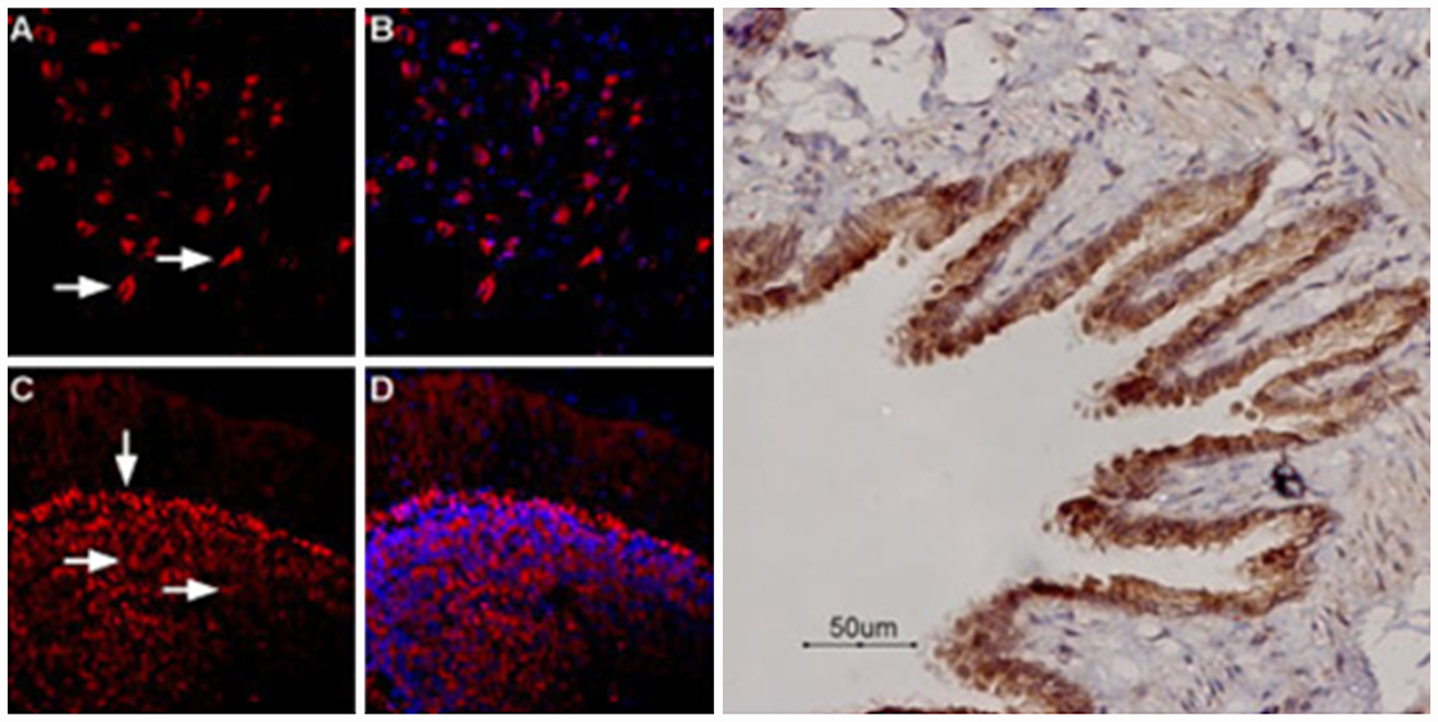
Using Anti-KCNN3 (KCa2.3, SK3) (N-term) Antibody (#APC-025), and Anti-GIRK1 (Kir3.1) Antibody (#APC-005), the authors immunostained coronal VNO sections. Slices probed separately with either antibodies showed that both KCa2.3 (SK3) and GIRK1 were highly expressed in cell bodies and dendritic knobes - following a pattern similar to TRPC2 expression - as well as in dendritic shafts. Next, using fluorescently tagged Anti-KCNN3 (KCa2.3, SK3) (N-term) Antibody, the group probed mice transgenic for Sk3 (Sk3T/T, inducible SK3 mouse knockout strain), before and after doxycycline (DOX) treatment. As expected, Sk3T/T VNO slices before DXO treatment showed normal expression pattern of SK3, in contrast to slices examined after DXO was applied, which had no detectable SK3 signals (Figure 1). Similarly, VNO slices of Girk1-/- stained with Anti-Kir3.1 (GIRK1) Antibody were free of GIRK1 fluorescence signal (Figure 2). Upon urine application, tertiapin-Q (a GIRK1 specific blocker) increased the inward current of VNO neurons from DXO-fed Sk3T/T mice (but had no effect on matched Girk1-/- cells), whereas apamin (a specific SK channels blocker) increased the current registered of VNO neurons from Girk1-/- (but had no effect on matched Sk3-/- cells).
Figure 1. Expression of KCa2.3 (SK3) in mouse VNO neuronsImmunohistochemical staining of in VNO sections from Sk3T/T mice fed with (right panel) or without (left panel) DOX diet using Anti-KCNN3 (KCa2.3, SK3) (N-term) Antibody (#APC-025).Adapted from Kim, S. et al. (2012) with permission of Nature America.
Lastly, the authors devised a finely tuned perfusion system, to address the discrepancies of the abovementioned outward current readings from VNO slices, as opposed to intact tissue preparations. Apparently, this inconsistency arises due to high and low K+ concentrations in the extracellular environment of the dendrite and cell body, respectively. Indeed, urine-rich stimulation of dendrites increased their firing rate, whereas diluted urine extracellular solutions attenuated it.
Figure 2. Expression of GIRK1 in mouse VNO neuronsImmunohistochemical staining of wild-type (left panel) and Girk1–/– (right panel) VNO neurons using Anti-GIRK1 (Kir3.1) Antibody (#APC-005).Adapted from Kim, S. et al. (2012) with permission of Nature America.
Wheeler D.G. et al. (2012) Cell 149, 1112.
Activity-dependent gene expression triggered by Ca2+ entry into neurons is critical for long-term changes in neural function, yet much remains unknown about how Ca2+ channels activate nuclear transcription factors.
A central issue in calcium signaling is how cells use Ca2+ delivered via multiple routes to trigger responses. A classic view is that multiple Ca2+ sources contribute convergently to the bulk cytoplasmic Ca2+ pool. An alternative view is that individual Ca2+ delivery systems may trigger specific cellular effects by using ‘‘private lines’’ of communication.
In the presented work, it was shown that two major classes of Ca2+ channels, CaV1 (L-type) and CaV2 (N-and P/Q-type) are involved in coupling membrane depolarization to cAMP response element-binding protein (CREB) phosphorylation and gene expression.
The study showed that there are differences between CaV1 and CaV2 in cell signaling mechanisms. In cultured rat SCG neurons, CaV2 channels were found to make up the majority of somatodendritic Ca2+ channels but appear less important than CaV1 in signaling to the nucleus. CaV1 channels are advantaged in their voltage-dependent gating and use nanodomain Ca2+ to drive local Ca2+/calmodulin-dependent protein kinase II (CaMKII) aggregation and trigger communication with the nucleus. In contrast, CaV2 channels must elevate [Ca2+]i microns away and promote CaMKII aggregation at CaV1 channels. These results were obtained in part by using Anti-CaV1.3 (CACNA1D) Antibody (#ACC-005) and Anti-CACNA1A (CaV2.1) Antibody (#ACC-001) in immunocytochemical staining.
Further, they examined the distribution of CaV1 and CaV2 channels relative to the endoplasmic reticulum (ER). Immunostaining of CaV2.1 channels (using #ACC-001) were found in large clusters, whereas CaV1.3 channels (using #ACC-005) were more diffusely distributed with smaller and less intense puncta. Data also show that ER and mitochondria selectively buffer Ca2+ entering through CaV2 channels and thereby restrict the impact of CaV2 channels on transcription.
These data suggest that the preference for Ca2+ entry mediated by CaV2 channels likely arises from their close relationship with sarco endoplasmic reticulum Ca2+ ATPase (SERCA) pumps in the ER. They found that the ER much more efficiently sequestered Ca2+ from CaV2 than from CaV1; and blockade of ER Ca2+ uptake mimics and occludes the effect of inhibiting mitochondrial Ca2+ uptake.
In conclusion, this study uncovers fundamental differences in the way that Ca2+ ions entering the cell through CaV1 versus CaV2 channels activate transcription. CaV1-derived Ca2+ acts locally, interacting with signaling machinery in the immediate vicinity of the channel to couple membrane depolarization to gene expression. In contrast, high-density CaV2 channel clusters generate large local elevations of [Ca2+]i in proximity to SERCA pumps, leading to preferential uptake by ER and mitochondria.
Arizono, M. et al. (2012) Sci. Signal. 5, ra27.
Astrocytes were for long believed to be important for extracellular ion homeostasis and nutrient support to neurons. Ca2+ imaging studies in astrocytes have strongly suggested that these star-like shaped cells play a more prominent role in brain function.
Past studies demonstrated that mGluR5 plays an important role regarding Ca2+ signaling in astrocytes. In an elegant study, Arizono, M. et al. explored the detailed subcellular Ca2+ dynamics of astrocytes and the spatial regulation of mGluR5-dependent signaling in these cells. Immunocytochemical staining of rat astrocytes using Anti-mGluR5 (extracellular) Antibody (#AGC-007) showed that the receptor is enriched at astrocyte processes rather than at somas, in accordance with Ca2+ imaging experiments which demonstrated that astrocytic processes are more sensitive to mGluR stimulation than the soma.
The lateral movement of mGluR5 in astrocytes was monitored using quantum dot-based single-particle tracking (QD-SPT). The procedure requires targeting streptavidin-conjugated nanoparticles of semi-conductive material to the antibody after it is incubated with the cells, thereby tracking the diffusion of probed mGluR5 in real-time by principles of optical relay, and eventually yielding movement trajectory and other important, often overlooked parameters. Ultimately, QD-SPT allowed the identification of an elusive mGluR5 membrane barrier which prevents the receptor from assimilating into the plasma membrane of the soma. In accordance with these results, disruption of the barrier resulted in disrupted Ca2+ signaling which was caused by a freely distributed mGluR5 receptor. In addition, mGluR5 localized to the soma was more static than mGluR5 in processes. Anti-P2X7 Receptor (extracellular) Antibody (#APR-008) was used to show that the barrier is specific to mGluR5 since in QD-SPT, P2X7 receptors were able to freely diffuse between the soma and the processes. The mGluR5 specific barrier may be important and crucial for local as opposed to global Ca2+ signaling which could be toxic under certain disease conditions.
Wydeven, N. et al. (2012) Proc. Natl. Acad. Sci. U.S.A. 109, 21492.
GIRK (G-protein gated inwardly rectifying K+) channels play a role in the inhibitory effect some neurotransmitters have on excitable cells. In neurons, GIRK2 homomers are largely responsible for the G-protein dependent gating, K+ selectivity and inward rectification. GIRK1 channels are also expressed in neurons. Abolishing GIRK1 expression, nearly eradicates GIRK-dependent signaling, strongly suggesting that GIRK1 potentiates GIRK2 channel activity, since there are no differences in GIRK2 membrane expression. In an attempt to map the areas of GIRK1 responsible for channel current potentiation, mutagenesis studies revealed that residues in the distal C-terminal tail and in the P-loop are important for receptor-induced activity and receptor-dependent/independent channel activity respectively. The mutant GIRK1 channels generated were expressed to wild type levels, and like GIRK1-/- cells, they had no effect on GIRK2 protein membrane expression as shown in western blot analysis using Alomone Labs Anti-GIRK2 (Kir3.2) Antibody (#APC-006).
Lv, P. et al. (2012) J. Neurosci. 32, 16314.
Afferent neurons from the spiral ganglion make up 95% of the total sensory cells. They are responsible for transmitting information from hair cells to the coclear nucleus. It is known that Ca2+ currents play an important role but little information is available about the properties of Ca2+ currents in posthearing SGNs due to a lack of electrophysiological recordings. Overcoming the technical constraints in isolating posthearing neurons from the inner ear enabled the identification of a plethora of CaV channels in that area. CaV2.2 was identified by using Alomone Labs Anti-CACNA1B (CaV2.2) Antibody (#ACC-002) and ω-Conotoxin MVIIA (#C-670), a known blocker of CaV2.2 evoked Ca2+ currents. CaV1.3 was detected with Anti-CaV1.3 (CACNA1D) Antibody (#ACC-005) and Nimodipine (#N-150), an antagonist of CaV1.3 Ca2+ currents. P/Q-type channels were detected with Anti-CACNA1A (CaV2.1) Antibody (#ACC-001) and ω-Agatoxin IVA (#STA-500) peptide toxin. Finally, SNX-482 (#RTS-500) was used to block R-type Ca2+ channels. This tedious study definitely sets the ground for Ca2+-dependent events in posthearing SGNs.
Nakaya, N. et al. (2012) J. Biol. Chem. 287, 37171.
Olfactomedin 1 (Olfm1) is a secreted glycoprotein detected in tissues of the central and peripheral nervous systems. In DRGs from embryonic and postnatal mice, Olfm1 is coexpressed with Nogo A receptor (NgR1), as detected in immunohistochemical staining using Alomone Labs Anti-Nogo Receptor (extracellular) Antibody (#ANT-008). Olfm1 binds to and inhibits signaling from the receptor by interfering with the interaction of NgR1 and p75NTR and LINGO-1. Through NgR1 binding, Olfm1 modulates axonal growth.
Bagher, P. et al. (2012) Proc. Natl. Acad. Sci. U.S.A. 109, 18174.
In arterioles, vasodilation is caused by the activation of Ca2+-activated KCa3.1 K+ channels which causes a hyperpolarization through nearby smooth muscle cells via KCa2.3 channels. The generation of Ca2+ responsible for activating KCa3.1 is caused by the activation of TRPV4 channel via low intraluminal pressure. The overall effect of the vasodilation reduces arterial tone thereby regulating blood flow. Immunohistochemical staining of pressurized arterioles was done using Alomone Labs Anti-KCNN4 (KCa3.1, SK4) Antibody (#APC-064) and Mouse Anti-KCNN4 (KCa3.1, SK4) (extracellular) Antibody (#ALM-051) in order to validate the expression of KCa3.1 in endothelial cells. Anti-KCNN3 (KCa2.3, SK3) (N-term) Antibody (#APC-025) was used to verify the expression of KCa2.3 in smooth muscle cells.
Liu, P. et al. (2012) J. Neurophysiol. 107, 3155.
In an attempt to study the contribution of KV3 channels to the repolarization of action potentials, it was found that Alomone Labs BDS-I (#B-400*), a known peptide blocker of KV3 channels, surprisingly produces broadening of the spike and accelerates the upstroke of the action potential. BDS-I does so by modulating voltage-gated Na+ channels. It enhances TTX-sensitive Na+ channels (highly effective on NaV 1.7 channels), and weakly inhibits TTX-resistant NaV channels.
*This product is discontinued and has been replaced by #STB-400.
Woo, D.H. et al. (2012) Cell 151, 25.
Astrocytes release glutamate via GPCRs in order to modulate synaptic transmission. Two newly identified mechanisms regulate glutamate release from astrocytes; one of which is TREK-1-dependent. It requires the activation of Gαi, dissociation of Gβγ, followed by the opening of the glutamate permeable KCNK2 (TREK-1) K+ channel through its N-terminal interaction with Gβγ. Immunohistochemistry electron microscopy using Alomone Labs Anti-KCNK2 (TREK-1) Antibody (APC-047) shows that the K+ channel tends to be expressed at the surface of the cell body and processes of astrocytes.
Ye, L. et al. (2012) Cell 151, 96.
Using a small molecule high troughput screen, Ye, L. et al. found that TRPV4 inhibition induces the expression of PGC1α in adipocytes, a key regulator of oxidative metabolism and thermogenesis. In addition, TRPV4 knockout mice, for which the lack of TRPV4 protein was verified using Alomone Labs Anti-TRPV4 Antibody (#ACC-034), or wild type mice treated with TRPV4 antagonists have increased thermogenesis in adipose tissues and do not develop diet-induced obesity, adipose inflammation and insulin resistance. The overall results of the paper may target TRPV4 for treating obesity and other metabolic diseases.
Okuno, H. et al. (2012) Cell 149, 886.
Using Anti-GluR1 (GluA1) (extracellular) Antibody (#AGC-004) in living hippocampal neurons, the authors examined GluR1 surface expression in correlation with the synaptic localization of the neuronal cytoskeleton-associated protein Arc (also called Arg3.1), a promising memory regulatory gene.
Dieni, S. et al. (2012) J. Cell Biol. 196, 775.
Using Anti-proBDNF Antibody (#ANT-006) in an array of experiments including immunofluorescence and immune electron microscopy in both wild type and BDNF knockout mice, the authors conclude that BDNF and its pro peptide are stored in large dense core vesicles in excitatory presynaptic neuronal terminals, suggesting an anterograde mode of action of BDNF, contrasting with the long-established retrograde model.