
A look through recent research that puts our directly conjugated antibodies to great use.
Flow cytometry is used around the world, every day. But we all know that, despite being incredibly powerful and useful, it’s riddled with challenges – from cell count and quality at the start, background and signal issues in the middle, and data analysis at the end. One of the keys to making it simpler and more effective is with antibodies directly conjugated to fluorophores. This direct conjugation not only eliminates the need to for extra steps labeling with a secondary antibody, but gives more precise results with less background.
Designing directly conjugated antibodies and panels for cytometry – all in-house – is something we’ve been doing for years. So, we wanted to explore some recent papers where these directly conjugated antibodies have been a huge help in flow cytometry. We’ll explore their role across different areas to highlight their versatility and reliability of these tools in contemporary research.
Illuminating Neutrophil Function
Let’s start with a paper from Molecular Nutrition and Food Research that explored the intriguing role of the TRPV1 channel in human neutrophils. They focused on how [6]-gingerol, a compound from ginger, affects these immune cells.1
The study revealed a notable upregulation of surface markers CD11b, CD66b, and the FPR1 receptor in neutrophils treated with [6]-gingerol. Intriguingly, these cells exhibited a 30% increase in CXCL8 secretion and ROS production, which suggests a strengthened immune response. Crucially, blocking TRPV1 negated these effects, underscoring the channel’s functional presence in neutrophils.
Here, our Anti-Human TRPV1 (extracellular)-FITC Antibody (#ACC-334-F) was instrumental in demonstrating TRPV1 expression on neutrophil surfaces. Through detailed flow cytometry, it allowed for a clear distinction between TRPV1 expression and isotype controls, as well as across samples from different donors.
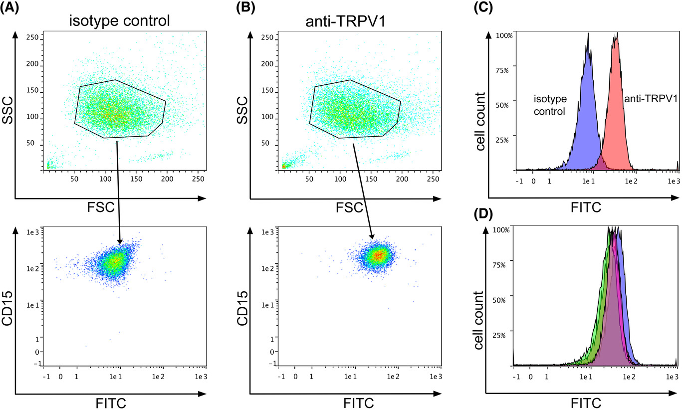
Figure 1. “The TRPV1 channel is expressed on the surface of human neutrophils. Flow cytometry of isolated human neutrophils, stained either with a TRPV1 specific antibody B) or the corresponding isotype control A), both conjugated to FITC. Cells were additionally stained for CD15. Numbers in the plots indicate either values for forward and side scatter or mean fluorescence intensity of the staining. C) Histogram overlay of the stains for TRPV1 and isotype control. D) Histogram overlay of the stains for TRPV1 of four individual donors.”
Deciphering Neurovascular Interactions in Brain Regeneration
In a paper from Cell Reports, researchers explored the interplay between neural stem cells (NSCs) and endothelial cells (ECs) in the brain’s subventricular zone (SVZ), a key area for adult brain regeneration, with a focus on Connexin 43 (Cx43).2
A core finding was the significant expression of Cx43 in both NSCs and ECs within the SVZ. The study revealed that deletion of Cx43 in these cells increases NSC proliferation and neuroblast generation, pointing to Cx43’s role in maintaining NSC quiescence. Furthermore, the research highlighted how Cx43 influences NSC behavior through mechanisms beyond its channel function, involving its cytoplasmic tail and ERK signaling.
Here, they used our Anti-EAAT1 (GLAST) (extracellular)-FITC Antibody (#AGC-021-F) antibody for precise identification and isolation of NSCs and ECs from adult mouse brain SVZ. This work not only underscores the utility of directly fluorophore-conjugated antibodies in flow cytometry but also advances our understanding of neurogenesis and potential therapies for neurodegenerative diseases.
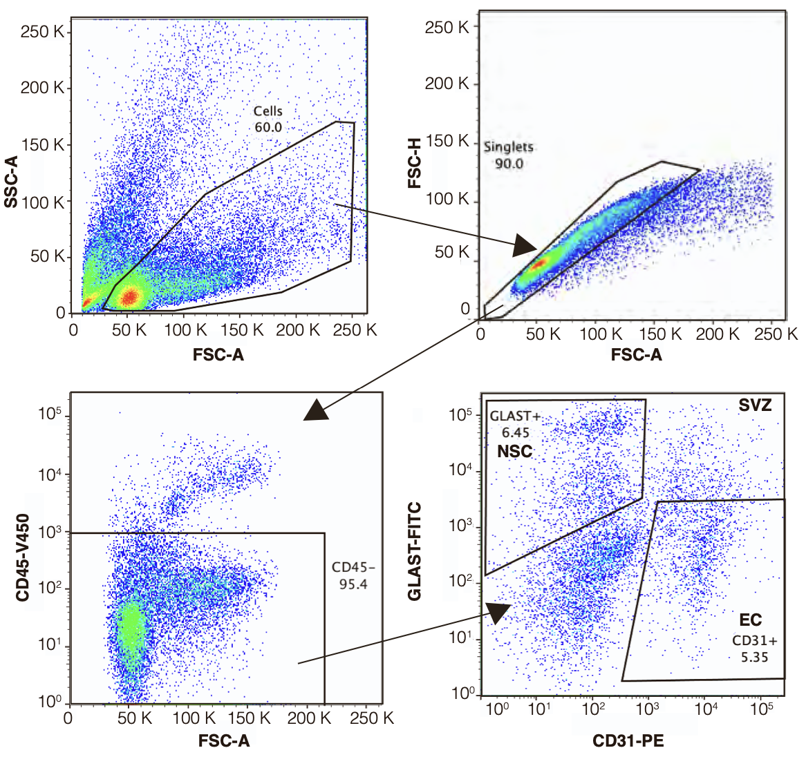
Figure 2. “Representative FACS plot showing the gating strategy for ECs and NSCs isolation from adult mouse brain SVZ. After excluding debris (top left panel) and doublets (top right panel), CD45- cells are selected (bottom left panel) and CD31+Glast- EC and CD31-Glast+ NSC are collected (bottom right panel). Percentages refer to the population of cells in the previous parent gate. Plots show 100,000 events. SSA: Side- scatter area, FSC: forward scatter area, FSH: forward-scatter height.”
Dexamethasone’s Role in COVID-19: A Flow Cytometry Study
Moving on to COVID-19 in Frontiers in Immunology, this significant research effort explored how dexamethasone affects immune response.3 This study helps to boost our understanding of the drug’s impact on inflammatory cytokine release and immune cell function.
Central to the study was the analysis of peripheral blood mononuclear cells (PBMCs) from COVID-19 patients, with and without dexamethasone treatment. Flow cytometry, using our Mouse Anti-Human Orai1 (extracellular)-ATTO Fluor-633 Antibody (#ALM-025-FR) revealed that dexamethasone reduces Kv1.3 potassium channel abundance in T cells and CD56dimNK cells, correlating with a decreased inflammatory response.
This research provides valuable insights into the mechanisms of dexamethasone’s effectiveness in treating severe COVID-19, emphasizing the role of flow cytometry to help us unravel the disease pathology and identify potential treatment responses.
Unraveling Oligodendrocyte Progenitor Development in the Rat Brain
This next research from Glia provides insights into brain development. It focuses on the p75 neurotrophin receptor (p75NTR) in oligodendrocyte progenitor cells (OPCs) within the subventricular zone (SVZ) of the postnatal rat brain.4 Here we saw our Anti-p75 NGF Receptor (extracellular)-ATTO Fluor-488 Antibody (#ANT-007-AG) in flow cytometry to analyze p75NTR expression in relation to other cell lineage markers. This approach showed that p75NTR is not only present but actively involved in OPC proliferation and differentiation, impacting early myelin formation.
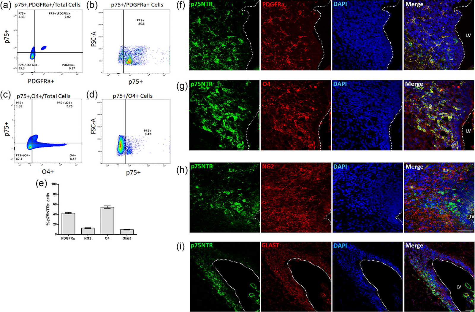
Figure 3. “p75NTR expressing cells of the dSVZ express OL lineage cell markers in vivo. SVZs from P7 rat pups were microdissected, dissociated, blocked with FcR blocker, stained for p75NTR, PDGFRα, O4, NG2 and GLAST and analyzed on a BD Fortessa flow cytometer. (a) Plot depicts cells double positive for p75NTR and PDGFRα out of the total live SVZ cell population. (b) Plot illustrates the percentage of p75NTR+ cells out of the PDGFRα+ subset. (c) Plot depicts cells double positive for p75NTR and O4 out of the total live SVZ cell population. (d) Plot illustrates the percentage of p75NTR+ cells out of the O4+ subset. Minimal coexpression was observed with NG2 or GLAST. Quad plots illustrate representative examples. (e) On average, 42% of the p75NTR expressing cells co-expressed PDGFRα+, 58% were O4+. NG2 and GLAST labeled 12.72% and 9.57% of the p75NTR population, respectively. n = 4 and error bars indicate mean ± SEM. Immunostaining of coronal sections from P7 WT rats confirms colocalization of p75NTR and (f) PDGFRα, (g) O4 and minimal colocalization between p75NTR and NG2 (h) or GLAST (i). Lateral ventricle labeled LV. Scale bars represents 50 μm.”
Targeting NGF-TrkA in Melanoma Immunosuppression
Finally, published this year in Nature Immunology, researchers have shed light on the role of the NGF-TrkA axis in melanoma immunosuppression.5 The research underscores how melanoma cells use the NGF–TrkA pathway to evade immune responses, highlighting an innovative approach to enhance immunotherapy effectiveness. In this paper, both Anti-TrkA (extracellular)-FITC Antibody (#ANT-018-F) and Anti-TrkA (extracellular)-ATTO Fluor-633 Antibody (#ANT-018-FR) were key in analyzing surface protein expression on T cells and tumor cells, further elucidating TrkA’s role in immune evasion and the potential of larotrectinib as a therapeutic agent.
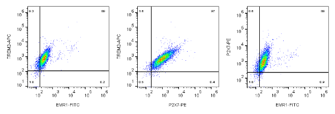
Microglia Exploration Panels
In addition to single directly conjugated antibodies, we have been working to bring you custom panels for flow cytometry. Just recently, we created a multicolor panel for cell surface markers in microglia, developed in mouse BV-2 microglia cells. This panel includes three key antibodies: Anti-EMR1 (ADGRE1) conjugated to FITC, Anti-P2X7 Receptor linked to PE, and Anti-TREM2 attached to APC. These antibodies enable the detailed analysis of microglial cell surface markers, providing valuable insights into the role and function of these cells in various neurological conditions. The flow cytometry data below, generated using this panel, show just how effective it is at identifying and characterizing microglia:
Conclusion
Hopefully you can appreciate the diverse applications of these directly fluorophore-conjugated antibodies in flow cytometry research. From investigating the immune responses of human neutrophils to [6]-gingerol, to exploring neurovascular interactions in brain regeneration, these antibodies have proven crucial. Also, their role in analyzing immune mechanisms in important diseases like COVID-19, oligodendrocyte progenitor development in the rat brain, and melanoma immunosuppression via the NGF-TrkA pathway, showcase not only their versatility but their importance in advancing scientific understanding across different fields of biological research.

References
- Andersen, G., Kahlenberg, K., Krautwurst, D. & Somoza, V. [6]-Gingerol Facilitates CXCL8 Secretion and ROS Production in Primary Human Neutrophils by Targeting the TRPV1 Channel. Mol. Nutr. Food Res. 67, 2200434 (2023).
- Genet, N. et al. Connexin 43-mediated neurovascular interactions regulate neurogenesis in the adult brain subventricular zone. Cell Rep. 42, 112371 (2023).
- Chimote, A. A. et al. Immune and ionic mechanisms mediating the effect of dexamethasone in severe COVID-19. Front. Immunol. 14, (2023).
- Joshi, S., Frondelli, M. J., Zanin, J. P., Levison, S. W. & Friedman, W. J. Oligodendrocyte progenitor development from the postnatal rat subventricular zone is regulated by the p75 neurotrophin receptor. Glia 71, 2383–2400 (2023).
- Yin, T. et al. Breaking NGF–TrkA immunosuppression in melanoma sensitizes immunotherapy for durable memory T cell protection. Nat. Immunol. 1–14 (2024) doi:10.1038/s41590-023-01723-7.