Immunohistochemistry (IHC) Protocols for Frozen Sections: Multiplex Staining
Target multiple proteins simultaneously with these multiplex IHC protocols using antibodies from the same or different host species.
Most immunohistochemistry (IHC) protocols describe experiments using only one antibody. In practice, however, you often need multiple antibodies against multiple targets. As the number of targets increases, so does the complexity of your multiplex experiment. However, with the right protocol, You can easily resolve the issues of managing antibodies from different host species.
Here, we describe our multiplex IHC protocol using both rabbit and guinea pig polyclonal primary antibodies on the same floating tissue sections.
If you have any problems, please see our extensive troubleshooting guides.
Preparation and Antigen Retrieval
- Rinse the floating sections with IHC phosphate-buffered saline (IHC-PBS) for 2 x 5 minutes.
| IHC-PBS (pH 7.4) | ||
|---|---|---|
| Reagent | Concentration | Volume/Weight |
| Na2HPO4 | 0.2 M | 80 ml |
| NaH2PO4 | 0.2 M | 16 ml |
| NaCl | 8 g | |
| Double distilled water | 860 ml | |
- For antigen retrieval and to quench endogenous peroxidase activity, incubate the floating sections with IHC-PBS with 0.2% hydrogen peroxide, 0.2% Triton X-100*, and 20% methanol, for 25 minutes at room temperature.
- Rinse the sections with IHC-PBS for 2 x 5 minutes.
*If your primary antibody targets an extracellular protein, reduce the Triton X-100 to 0.05% in both the primary and secondary antibody solutions.
Two options for multiplex IHC are available here:
A. Primary Antibodies Raised in Different Hosts
B. Primary Antibodies Raised in the Same Host
OPTION A: Primary Antibodies Raised in Different Hosts
- Incubate the sections with a cocktail containing two primary antibodies:
a. The first raised in rabbit (1:200 to 1:400)
b. The second raised in guinea pig (1:200 to 1:400)
Dilute both antibodies in Multiplex Antibody Solution for 1 hour at room temperature.
| Multiplex Antibody Solution | |
|---|---|
| Reagent | % of final volume |
| IHC-PBS | 95.65 |
| Triton X-100* | 0.3 |
| Tween-20 | 0.05 |
| Normal goat serum (NGS) | 2 |
| Normal donkey serum | 2 |
*If your primary antibody targets an extracellular protein, reduce the Triton X-100 to 0.05% in both the primary and secondary antibody solutions.
- Incubate the sections at 4°C overnight.
- Rinse the sections with IHC-PBS containing 2% NGS and 2% NDS for 2 x 5 minutes.
- Incubate the sections with a cocktail of diluted secondary antibodies:
Anti-rabbit conjugated to a fluorescent dye
Anti-guinea pig conjugated to a different fluorescent dye
Dilute both antibodies in Multiplex Antibody Solution for 1 hour at room temperature. - Incubate the sections at 4°C overnight.
- Proceed to Mounting and Detection.
OPTION B: Primary Antibodies Raised in the Same Host
- Incubate the sections with rabbit primary antibody (un-conjugated) diluted in Antibody Solution for 1 hour at room temperature.
| Antibody Solution | |
|---|---|
| Reagent | % of final volume |
| IHC-PBS | 97.65 |
| Triton X-100* | 0.3 |
| Tween-20 | 0.05 |
| Normal goat serum (NGS) | 2 |
*If your primary antibody targets an extracellular protein, reduce the Triton X-100 to 0.05% in both the primary and secondary antibody solutions.
- Incubate the sections at 4°C overnight.
- Rinse the sections with IHC-PBS containing 2% NGS for 2 x 5 minutes.
- Incubate the sections with the secondary antibody (goat anti-rabbit conjugated to a fluorescent dye) in Antibody Solution for 1 hour at room temperature.
- Incubate the sections at 4°C overnight.
- Rinse the sections with IHC-PBS containing 2% NGS for 2 x 5 minutes.
- Incubate the sections with 2% normal rabbit serum (NRS) (to saturate residual binding ability of the secondary goat anti-rabbit antibody), for 1 hour at room temperature.
- Rinse the sections with IHC-PBS containing 2% NGS for 2 x 5 minutes.
- Incubate the sections with rabbit primary antibody conjugated to a different fluorescent dye, diluted 1:50–1:60 in Antibody Solution for 1 hour at room temperature.
- Incubate the sections at 4°C overnight.
- Proceed to Mounting and Detection.
*If your primary antibody targets an extracellular protein, reduce the Triton X-100 to 0.05% in both the primary and secondary antibody solutions.
Mounting and Detection
- Rinse the sections with IHC-PBS (containing 2% NGS) for 2 x 5 minutes.
- Mount sections on slides and dry for 2 hours to overnight.
- Stain the mounted sections with DAPI stain (5 mg/ml stock solution in deionized water or dimethylformamide (DMF); dilute to 500 nM in IHC-PBS for final use) to label all cells in the field. Incubate in DAPI at room temperature for 5 minutes.
- Apply coverslips using the adhesive Immu-MountTM (ShandonTM).
- Detect with a microscope.
Example Data

Figure 1: Multiplex staining of calnexin and presenilin-1 in the mouse cortex, using two rabbit primary antibodies. The sections were incubated with a cocktail of rabbit Anti-Calnexin conjugated with ATTO red (ACS-009-AR) and rabbit Anti-Presenilin conjugated with ATTO green (AIP-011-AG) antibodies. A) Calnexin staining (red) appeared in the neuronal profiles. B) Presenilin-1 staining (green) in the same section appeared in the neuronal profiles and apical dendrites (arrows). C) A merged image of A and B demonstrated the colocalization of calnexin and presenilin-1 in several neurons (arrows). The cell nuclei are stained with DAPI (blue) as the counterstain.
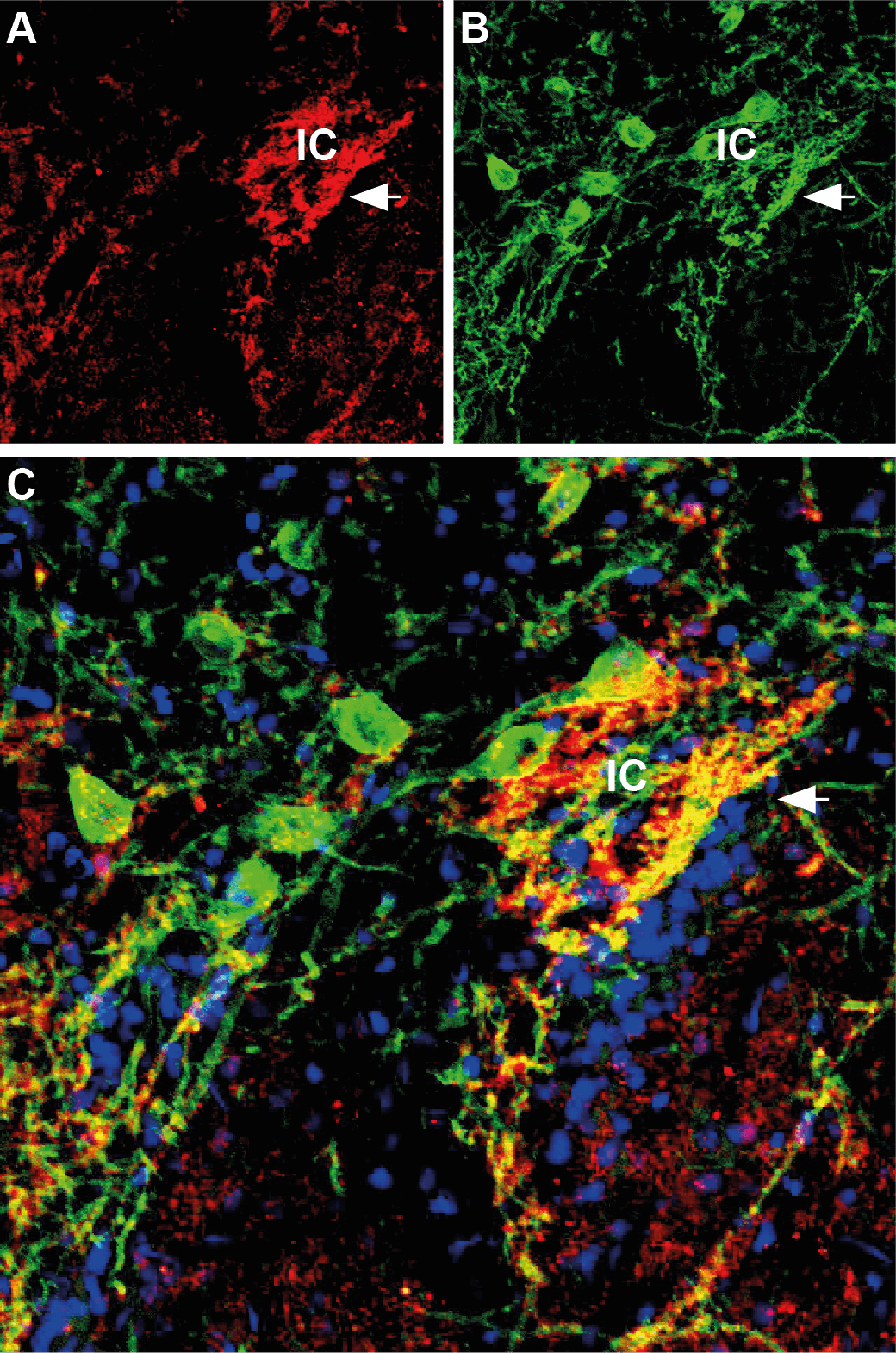
Figure 2: Multiplex staining of synapsin II (SYN2) and GABA(A) α1 receptor in the rat striatum using primary antibodies from different hosts. The sections were incubated with a cocktail of rabbit Anti-Synapsin II (ANR-015) and guinea pig Anti-GABA(A) α1 receptor (AGP-083) antibodies, followed by incubation with donkey anti-rabbit Cy3 (red) and goat anti-guinea pig Alexa 488 (green). A) Synapsin II (red) appeared in the Islands of Calleja (arrow). B) The GABA(A) α1 receptor (green) was detected in the Islands of Calleja. C) A merged image of A and B demonstrated partial colocalization of synapsin II and GABA(A) α1 receptor. The cell nuclei are stained with DAPI (blue).