Overview
- Peptide KKGWMDPQSKGIQTGRC, corresponding to amino acid residues 136-152 of mouse P2X7 receptor (Accession Q9Z1M0). Extracellular loop.
 Cell surface detection of P2RX7 in intact living human THP-1 monocytic leukemia cells:___ Cells.
___ Cells + Rabbit IgG isotype control-APC (#RIC-001-FR).
___ Cells + P2X7 Receptor (extracellular)-ATTO Fluor-633 (#APR-008-FR), (5µg).
- Rat brain glioma (C6) cells.
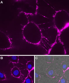 Expression of P2RX7 in rat brain glioma (C6) cellsCell surface detection of P2RX7 in intact living rat C6 cells. A. Extracellular staining of cells with Anti-P2X7 Receptor (extracellular)-ATTO Fluor-633 Antibody (#APR-008-FR), (1:25, purple). B. Merged image of A and nuclear staining using DAPI as the counterstain (blue). C. Merged image of B with cells live image.
The P2X7 receptor is a member of the ionotropic P2X receptor family that is activated by ATP. To date, this family is composed of seven cloned receptor subtypes, named P2X1-P2X7.
The different P2X receptors show distinct expression patterns. P2X1-6 have been found in the central and peripheral nervous system, while the P2X7 receptor is found in cells of the immune system, particularly antigen presenting cells, and microglia. The P2X7 receptor mediates the release of proinflamatory cytokines, stimulation of transcription factors and may also have an important role in apoptosis.1-3
Different techniques have been used to characterize the P2X7 receptor. Most of them investigated pores, ion channels (electrophysiology) and membrane alterations (calcium microfluorometry, dye uptake, membrane depolarization and ion influx analysis). With the introduction of flow cytometry, it is now possible to analyze multiple cell parameters such as cell cycle, cell membrane alteration, calcium influx and cell phenotype.4
