Overview
- Peptide KKGWMDPQSKGIQTGRC, corresponding to amino acid residues 136-152 of mouse P2X7 receptor (Accession Q9Z1M0). Extracellular loop.

 Cell surface detection of P2RX7 in live intact mouse BV-2 microglia cells:___ Cells.
___ Cells + rabbit IgG isotype control-FITC.
___ Cells + Anti-P2X7 Receptor (extracellular)-FITC Antibody (#APR-008-F), (5 µg). Cell surface detection of P2RX7 in live intact human THP-1 monocytic leukemia cells:___ Cells.
___ Cells + rabbit IgG isotype control-FITC.
___ Cells + Anti-P2X7 Receptor (extracellular)-FITC Antibody (#APR-008-F), (5 µg).- The control antigen is not suitable for this application.
 Expression of P2RX7 in human K562 cellsCell surface detection of P2RX7 in living K562 cells stained with Anti-P2X7 Receptor (extracellular)-FITC Antibody (#APR-008-F).
- Ding, Y. et al. (2000) J. Auton. Nerv.Syst. 81, 289.
- Kim, M. et al. (2001) EMBO J. 20, 6347.
- Chizh, B.A. et al. (2000) Pharmacol. Rev. 53, 553.
- Nihei, O.K. et al. (2000) Mem.Inst.Oswaldo Cruz. 95, 415.
The P2X7 receptor is a member of the ionotropic P2X receptor family that is activated by ATP. To date, this family is composed of seven cloned receptor subtypes, named P2X1-P2X7.
The different P2X receptors show distinct expression patterns. P2X1-6 have been found in the central and peripheral nervous system, while the P2X7 receptor is found in cells of the immune system, particularly antigen presenting cells, and microglia. The P2X7 receptor mediates the release of proinflamatory cytokines, stimulation of transcription factors and may also have an important role in apoptosis.1-3
Different techniques have been used to characterize the P2X7 receptor. Most of them investigated pores, ion channels (electrophysiology) and membrane alterations (calcium microfluorometry, dye uptake, membrane depolarization and ion influx analysis). With the introduction of flow cytometry, it is now possible to analyze multiple cell parameters such as cell cycle, cell membrane alteration, calcium influx and cell phenotype.4
Anti-P2X7 Receptor (extracellular) Antibody (#APR-008) is a highly specific antibody directed against an epitope of the mouse protein. The antibody can be used in western blot, indirect flow cytometry, immunohistochemistry, live cell imaging, and immunocytochemistry applications. It has been designed to recognize P2X7 purinergic receptor from mouse, rat, and human samples.
Anti-P2X7 Receptor (extracellular)-FITC Antibody (#APR-008-F) is directly conjugated to fluorescein isothiocyanate (FITC). This labeled antibody can be used in immunofluorescent applications such as direct live cell flow cytometry and immunocytochemistry.
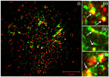
Multiplex staining of P2X7 receptor and cholera toxin in MC3T3-E1 cells.Immunocytochemical staining with labeled cholera toxin and Anti-P2X7 Receptor (extracellular)-FITC Antibody (#APR-008-F). The ganglioside GM1, the receptor for cholera toxin is concentrated in caveolae. Staining shows that P2X7 receptor colocalizes with cholera toxin.
Adapted from Gangadharan, V.et al. (2014) with permission of the American Physiological Society.
Applications
Citations
 Powered by Bioz
- Western blot analysis of mouse neutrophil lysate using #APR-008. Tested in P2X7-/- mice.
Karmakar, M. et al. (2016) Nat. Commun. 7, 10555.
- Human PBMCs.
Luchting, B. et al. (2016) J. Neuroinflammation 13, 1.
- Gangadharan, V. et al. (2014) Am. J. Physiol. 308, C41.
- Martel Gallegos, G. et al. (2010) Purinergic Signal. 6, 297.
- Milius, D. et al. (2008) Neurosci. Lett. 446, 45.
- Milius, D. et al. (2007) Toxicology 238, 60.
