Overview
- Peptide (C)DETSSRKEKWDLR, corresponding to amino acid residues 287-299 of rat prostanoid EP2 receptor (Accession Q62928). 3rd extracellular loop.

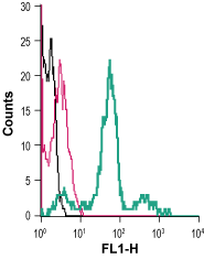
 Cell surface detection of PTGER2 in live intact human THP-1 monocytic leukemia cells:___ Cells.
___ Cells + rabbit IgG isotype control-FITC.
___ Cells + Anti-Prostaglandin E Receptor EP2/PTGER2 (extracellular)-FITC Antibody (#APR-064-F), (2.5 µg). Cell surface detection of PTGER2 in live intact mouse J774 macrophage cells:___ Cells.
___ Cells + rabbit IgG isotype control-FITC.
___ Cells + Anti-Prostaglandin E Receptor EP2/PTGER2 (extracellular)-FITC Antibody (#APR-064-F), (2.5 µg).
- Campbell, W.B. et al. (1990) The Pharmacological Basis of Therapeutic 8, 600.
- Davies, P. et al. (1992) Inflammation: Basic Principles and Clinical Correlates 2, 123.
- Coleman, R.A. et al. (1992) Comprehensive Medicinal Chemistry 3, 123.
- Ferreri, N.R. et al. (1992) J. Biol. Chem. 267, 9443.
- Bastien, L. et al. (1994) J. Biol. Chem. 269, 11673.
Prostaglandin E2 (PGE2) is involved in a number of physiological and pathophysiological events in many tissues throughout the body1. The physiological actions of PGE2 are mediated through its interaction with cell surface prostaglandin E receptors.
There are three pharmacologically defined subtypes of the EP receptor, EP1, EP2, and EP3, and these subtypes are suggested to be different in their signal transduction2. These receptors belong to the G-protein coupled receptor (GPCR) superfamily. Like all members they have seven transmembrane domains with an extracellular N-terminal tail and an intracellular C-terminus. The EP2 receptor is expressed in the vasculature, the gastrointestinal tract, kidney and also in the ciliary muscles of the eye3.
PGE2 is known to play a central role in the pathophysiology of inflammation in synergy with other proinflammatory mediators. PGE2 inhibits the function and the proliferation of T cells and the histamine release from mast cells by increasing the intracellular level of cAMP4.
The EP2 subtype is thought to be in part responsible for vasodilation, oedema formation, hyperanalgesia, modulation of the immune system, and the breakdown of bone and cartilage associated with disorders such as rheumatoid arthritis5.
Application key:
Species reactivity key:
Anti-Prostaglandin E Receptor EP2/PTGER2 (extracellular) Antibody (#APR-064) is a highly specific antibody directed against an epitope of the rat protein. The antibody can be used in western blot, immunohistochemistry, and indirect flow cytometry applications. It has been designed to recognize EP2 receptor from rat, mouse, and human samples.
Anti-Prostaglandin E Receptor EP2/PTGER2 (extracellular)-FITC Antibody (#APR-064-F) is directly conjugated to fluorescein isothiocyanate (FITC). The antibody can be used in immunofluorescent applications such as direct live cell flow cytometry.
