Overview
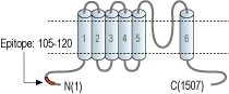
- Peptide (C)HTFQGKEWDPKKHVQE, corresponding to amino acid residues 105-120 of mouse TRPM2 (Accession Q91YD4). Intracellular, N-terminus.
 Western blot analysis of rat lung membrane (lanes 1 and 4), rat brain membrane (lanes 2 and 5) and human K562 chronic myelogeneous cells (lanes 3 and 6) lysates:1-3. Anti-TRPM2 Antibody (#ACC-043), (1:200).
4-6. Anti-TRPM2 Antibody, preincubated with TRPM2 Blocking Peptide (#BLP-CC043).
 Expression of TRPM2 in rat lungImmunohistochemical staining of rat lung paraffin embedded sections using Anti-TRPM2 Antibody (#ACC-043), (1:100). TRPM2 is expressed in the respiratory epithelium (black arrows). In addition, staining is also present in smooth muscle, both bronchiolar (arrowheads) and vascular (red arrows). Hematoxilin is used as the counterstain.
- Mouse brain endothelial cells (1:200), (Park, L. et al. (2014) Nat. Commun. 5, 5318.).
- Fleig, A. et al. (2004) Trends. Pharmacol. Sci. 25, 633.
- Kraft, R. et al. (2005) Pflugers. Arch. 451, 204.
- Huang, C.L. (2004) J. Am. Soc. Nephrol. 15, 1690.
- Montell, C. (2005) Sci. STKE. 2005, re3.
- Du, J. et al. (2009) Proc. Natl. Acad. Sci. U.S.A. 106, 7239.
The mammalian melastatin-related transient receptor potential (TRPM) is a subfamily of the TRP family. The family was named after the first discovered member, melastatin (TRPM1) whose gene was identified in metastatic and benign melanomas1-3.
The TRPM family consists of eight members designated as TRPM1-8 that can be further divided into four pairs: TRPM1 and TRPM3; TRPM2 and TRPM8; TRPM4 and TRPM5; and TRPM6 and TRPM71,2.
The TRPM proteins share structural homology with other members of the TRP superfamily channels; six putative transmembrane domains, and cytoplasmic N- and C-termini. However, due to their long N- and C-termini they are also named the long TRP channel family1-3.
The C-terminal regions of three TRPM members (TRPM2, TRPM6 and TRPM7) are occupied by enzymatic domains. Almost all the C-terminal region of TRPM2 has an enzymatic domain with sequence similarities to Nudix hydrolases (cleave mononucleotide and dinucleotide polyphosphates2,4.
TRPM2 (LTRpC2, TrpC7) is a Ca2+-permeable, nonselective cation channel that is predominantly expressed in various regions of the brain, where it is preferentially localized in microglial cells, the host macrophages of the central nervous system and is also expressed in other tissues, including spleen, heart, liver, lung, and bone marrow. Several splice variants of TRPM2 have been identified5,3.
Anti-TRPM2 Antibody (#ACC-043) is a highly specific antibody directed against an intracellular epitope of mouse TRPM2. The antibody can be used in western blot, immunocytochemistry, and immunohistochemistry applications. It has been designed to recognize TRPM2 from mouse, rat, and human samples.
Applications
Citations
 Powered by Bioz
- Western blot and immunocytochemistry of mouse brain endothelial cells. Tested in siRNA treated cells.
Park, L. et al. (2014) Nat. Commun. 5, 5318.
- Human ARPE-19 retinal pigment epithelial cell lysate (1:200).
Melendez Garcia, R. et al. (2016) Ebiomedicine 7, 35. - Mouse brain endothelial cell lysate (1:200).
Park, L. et al. (2014) Nat. Commun. 5, 5318.
- Human pulpal tissue sections (1:100).
Rowland, K.C. et al. (2007) J. Endod. 33, 245.
- Mouse brain endothelial cells (1:200).
Park, L. et al. (2014) Nat. Commun. 5, 5318.
