
Photo by Ravi Patel
Erythrocyte adenosine A2B receptor signaling cascade protects against age-related decline in cognition.
Global life expectancies continue to increase thanks to positive public health changes and the ever-evolving advances in modern medicine. But alongside this increase in life expectancy, we also see an increase in the number of people who experience age-related cognitive decline. A lot of the research into this decline has focused on gene expression changes in organs, but this fails to capture the complete picture, especially when it comes to aspects like hearing loss.
Instead of looking at organs, research published in PLOS Biology from Qiang et al. at The University of Texas McGovern Medical School turned its attention to erythrocytes and oxygen availability (1). Aging has long been suspected to go hand-in-hand with reduced oxygen delivery to tissues and the subsequent inflammatory response following macrophage activation and the release of proinflammatory chemokines and cytokines (2). This cascade is often referred to as “inflamm-aging” (3).
Since oxygen levels in the blood decline with age, the researchers began to think more about oxygen delivery to the brain. Adenosine signaling plays a role in brain function, and extracellular adenosine in particular – which increases in the brain with age – can initiate a hypoxic response by activating cell surface adenosine receptors. The four adenosine receptors activated during hypoxic responses, and which could have behavioral consequences, are adenosine A1 receptor (ADORA1) and adenosine A2A receptor (ADORA2A), both highly expressed in neurons and gliocytes. And adenosine A3 receptor (ADORA3) and adenosine A2B receptor (ADORA2B), which are found throughout the brain but a generally low level of expression.
Earlier work highlighted how ADORA2B was regulating metabolic adaptation to high-altitude hypoxia (4) through elevated adenosine levels. This ADORA2B signaling in turn induced AMP-activated protein kinase (AMPK) to activate bisphosphoglycerate mutase (BPGM) and drive 2,3-bisphosphoglycerate (2,3-BPG) synthesis – an allosteric modulator of hemoglobin that releases oxygen to counter the effects of hypoxia (5).
So, could aging and its effects on the body be naturally held off by a specific adenosine receptor? Bypassing the more commonly investigated cell types like immune cells and neurons, the researchers instead looked at erythrocytes – the only cell type delivering oxygen to the brain – which also express ADORA2B on their surface. To investigate this, they engineered an erythrocyte-specific ADORA2B knockout mouse model (eAdora2b−/−) to study the role of these adenosine receptors in hypoxia and aging.
Since the protein is on the surface, an ideal way of targeting that would be with something like our Anti-Adenosine A2B Receptor (extracellular) (AAR-003) Antibody, designed to target the extracellular region of ADORA2B.
In their knockout mice, they saw an early onset decline in cognitive and cochlear function. At the same time, young eAdora2b−/− mice experienced a substantial activation of innate immune cells and cytokines in the hippocampus, cortex, and cochlea. In experiments with bouts of hypoxia, mice were less able to deal with low oxygen levels due to the attenuation of HIF1α-induced targeted genes.
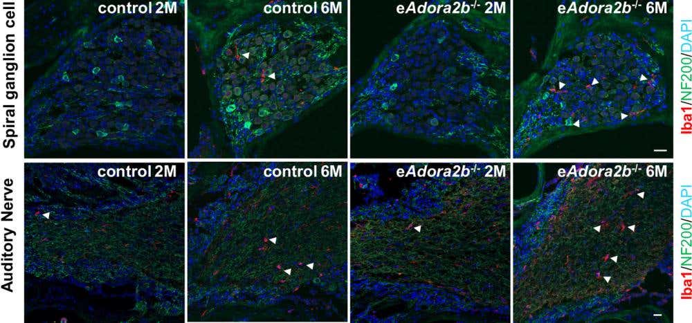
Figure 1. Increased abundance of enlarged macrophages and pro-inflammatory cytokines in the cochlea of eAdora2b−/− mice at 2 versus 6 months old. Representative images of macrophages around spiral ganglion cells and AN were identified by Iba1 and NF200 staining. Scale bar, 20 μm. Arrowheads indicate macrophage cells. Figure reproduced from Qiang, Q. et al. PLoS Biol. 19, (2021).
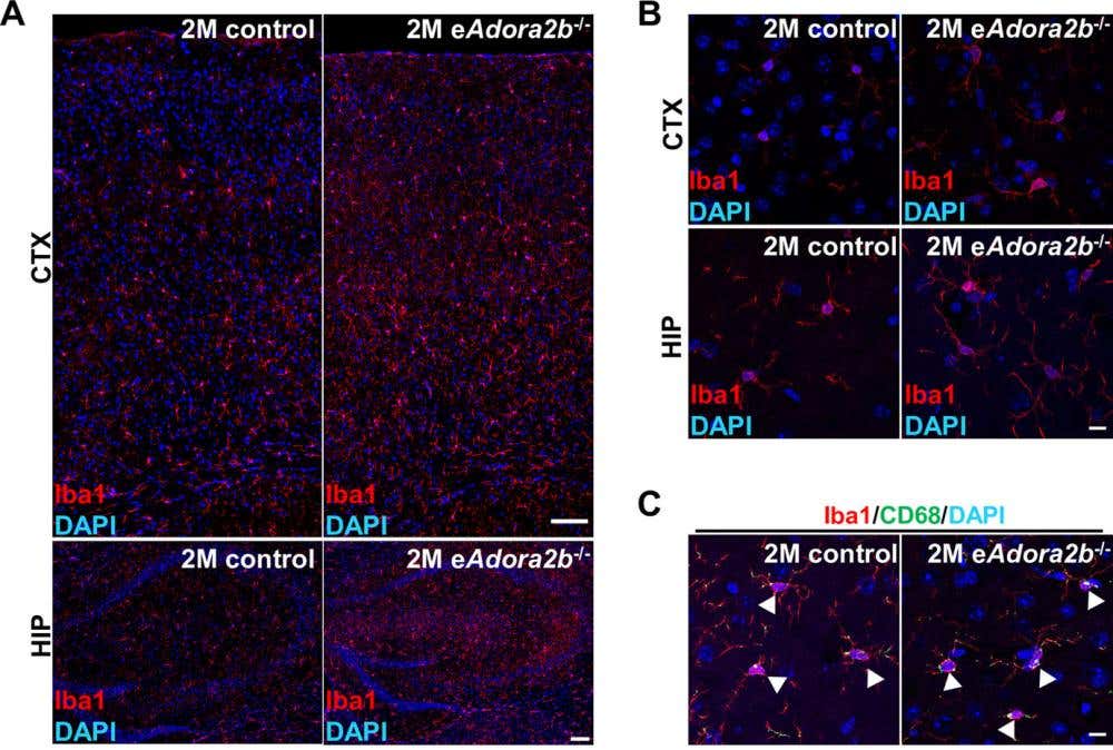
Figure 2. Increased abundance of activated microglia/macrophages and proinflammatory cytokines in tissues of eAdora2b−/− mice following hypoxia treatment. (A–C) Representative images of microglia from the brain. (A) Representative images of microglia visualized by Iba1 staining in CTX and HIP are shown. Scale bar, 100 μm. (B) Representative high-magnification images show the morphology of microglia in CTX and HIP. Scale bar, 10 μm. (C) Representative high-magnification images of activated microglia in the brain visualized by co-staining Iba1 and CD68. Scale bar, 10 μm. Arrowheads indicate areas of co-staining. CTX, cerebral cortex; HIP, hippocampus. Figure reproduced from Qiang, Q. et al. PLoS Biol. 19, (2021).
What the results here show is that erythrocyte ADORA2B signaling works to counteract hypoxia-exacerbated aging effects by protecting and maintaining normal levels of cognitive and cochlear function. Looking more at specific pathway involvement, the research team showed that it was erythrocyte-specific ADORA2B–AMPK–BPGM signaling axis activation affecting the decline in cognitive and cochlear function, as well as the inflammatory response, caused by aging and hypoxia.
The ADOAR2B activation of AMPK and BPGM in works as an essential component in “anti-aging” and “anti-hypoxia” through its induction of 2,3-BPG production and oxygen delivery, which slow age-related functional decline.
Dr. Xia, the study leader, said “Red blood cells have an irreplaceable function to deliver oxygen to maintain bioenergetics of every single cell within our body. However, their function in age-related cognition and hearing function remains largely unknown. Our findings reveal that the red blood cell ADORA2B signaling cascade combats the early onset of age-related decline in cognition, memory, and hearing by promoting oxygen delivery in mice and immediately highlight multiple new rejuvenating targets”.
This work opens the possibility of targeting this axis for therapeutic purposes, but more research will need to be done into purinergic signaling to understand the broader picture and other important pathway members.


 Antibody")
