-
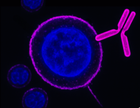
 A Rabbit Polyclonal Antibody to Human TRPV6 Channel
- Product Code: ACC-028
- Short Description: A Rabbit Polyclonal Antibody to Human TRPV6 Channel
- Host: Rabbit
- Label: Unconjugated
- Reactivity: Human
- Applications: Live cell imaging, Western blot, Immunocytochemistry, Immunofluorescence
- Target: Epithelial calcium channel 2, ECaC2, Calcium transport protein 1, CaT1, CaT-like, CaT-L, Transient receptor potential cation channel subfamily V member 6
-

 A Rabbit Polyclonal Antibody to CACNA2D1 (CaVα2δ1 Subunit)
- Product Code: ACC-015
- Short Description: A Rabbit Polyclonal Antibody to CACNA2D1 (CaVα2δ1 Subunit)
- Host: Rabbit
- Label: Unconjugated
- Reactivity: Human, Mouse
- Applications: Live cell imaging, Western blot, Immunocytochemistry, Immunofluorescence
- Target: Voltage-dependent Ca2+ channel subunit alpha-2/delta-1, L-type calcium channel alpha 2 polypeptide, Dihydropyridine-sensitive L-type calcium channel alpha-2/delta subunit
-
 A Rabbit Polyclonal antibody to BMPR1A (extracellular)
- Product Code: ABR-043
- Short Description: A Rabbit Polyclonal antibody to BMPR1A (extracellular)
- Host: Rabbit
- Label: Unconjugated
- Reactivity: Human, Rat, Mouse
- Applications: Indirect flow cytometry, Live cell imaging, Western blot, Immunohistochemistry
- Target: Bone Morphogenetic Protein Receptor Type-1A, Activin Receptor-Like Kinase 3, ALK3, Serine/Threonine-Protein Kinase Receptor R5, SKR5, CD292
-
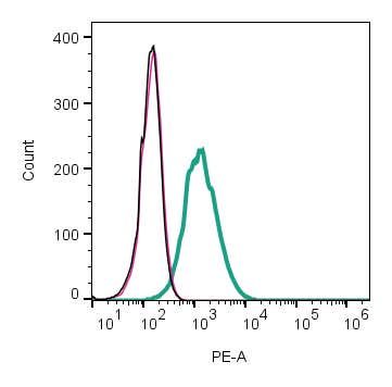 A Rabbit Polyclonal Antibody to ACVRL1
- Product Code: ABR-041
- Short Description: A Rabbit Polyclonal Antibody to ACVRL1
- Host: Rabbit
- Label: Unconjugated
- Reactivity: Rat, Mouse
- Applications: Indirect flow cytometry, Live cell imaging, Western blot
- Target: Activin A Receptor Like Type 1, ALK1, Serine/Threonine-Protein Kinase Receptor R3, SKR3, TGF-B Superfamily Receptor Type I, TSR-I
-
 A Rabbit Polyclonal Antibody to BAI2
- Product Code: ABR-022
- Short Description: A Rabbit Polyclonal Antibody to BAI2
- Host: Rabbit
- Label: Unconjugated
- Reactivity: Human, Rat, Mouse
- Applications: Western blot
- Target: Brain-specific angiogenesis inhibitor 2, Adhesion G protein-coupled receptor B2, ADGRB2
-
 A Rabbit Polyclonal Antibody to BAI1 Conjugated to the Fluorescent Dye FITC
- Product Code: ABR-021-F
- Short Description: A Rabbit Polyclonal Antibody to BAI1 Conjugated to the Fluorescent Dye FITC
- Host: Rabbit
- Label: FITC
- Reactivity: Human, Rat, Mouse
- Applications: Live cell imaging, Flow cytometry
- Target: Brain-specific angiogenesis inhibitor 1, Adhesion G protein-coupled receptor B1, ADGRB1, GDAIF
-
 A Rabbit Polyclonal Antibody to BAI1
- Product Code: ABR-021
- Short Description: A Rabbit Polyclonal Antibody to BAI1
- Host: Rabbit
- Label: Unconjugated
- Reactivity: Human, Rat, Mouse
- Applications: Indirect flow cytometry, Live cell imaging, Western blot, Immunofluorescence, Immunohistochemistry
- Target: Brain-specific angiogenesis inhibitor 1, Adhesion G protein-coupled receptor B1, ADGRB1, GDAIF
-
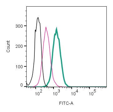 A Rabbit Polyclonal antibody to Human BDKRB1(extracellular) conjugated to FITC fluorophore.
- Product Code: ABR-013-F
- Short Description: A Rabbit Polyclonal antibody to Human BDKRB1(extracellular) conjugated to FITC fluorophore.
- Host: Rabbit
- Label: FITC
- Reactivity: Rat, Mouse
- Applications: Live cell imaging, Flow cytometry
- Target: BK-1 receptor, BKRB1, B1R, BKR2
-
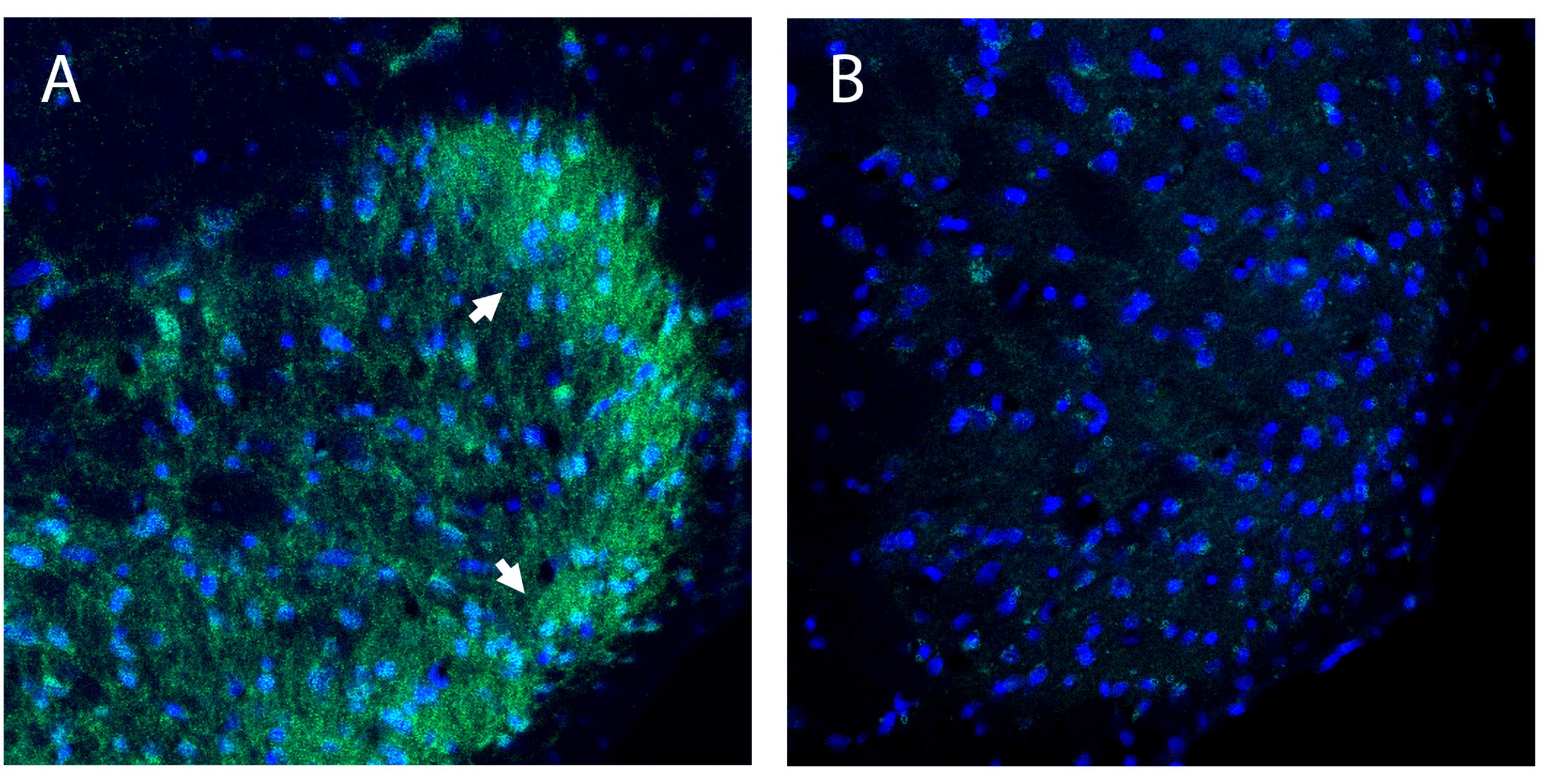 A Rabbit Polyclonal Antibody to B1 Bradykinin Receptor
- Product Code: ABR-013
- Short Description: A Rabbit Polyclonal Antibody to B1 Bradykinin Receptor
- Host: Rabbit
- Label: Unconjugated
- Reactivity: Rat, Mouse
- Applications: Indirect flow cytometry, Live cell imaging, Western blot
- Target: BK-1 receptor, BKRB1, B1R, BKR1
-
 A Rabbit Polyclonal Antibody to Bombesin Receptor 3
- Product Code: ABR-003
- Short Description: A Rabbit Polyclonal Antibody to Bombesin Receptor 3
- Host: Rabbit
- Label: Unconjugated
- Reactivity: Human, Rat, Mouse
- Applications: Indirect flow cytometry, Live cell imaging, Western blot, Immunohistochemistry
- Target: BB3R, BRS3
Loading...