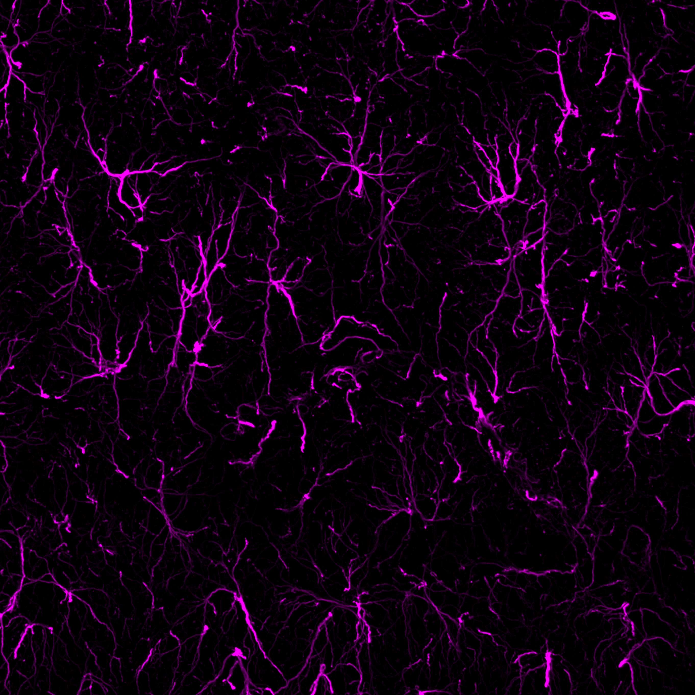
-
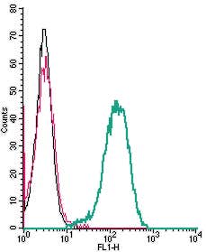 A Rabbit Polyclonal Antibody to C3a Anaphylatoxin Receptor
- Product Code: AAR-031
- Short Description: A Rabbit Polyclonal Antibody to C3a Anaphylatoxin Receptor
- Host: Rabbit
- Label: Unconjugated
- Reactivity: Human
- Applications: Indirect flow cytometry, Live cell imaging, Western blot
- Target: C3a anaphylatoxin chemotactic receptor, C3a anaphylatoxin receptor, Complement component 3a receptor 1, C3a receptor, C3AR1, C3R1, AZ3B, HNFAG09
-
 A Rabbit Polyclonal Antibody to CX3CR1 Chemokine Receptor
- Product Code: ACR-059
- Short Description: A Rabbit Polyclonal Antibody to CX3CR1 Chemokine Receptor
- Host: Rabbit
- Label: Unconjugated
- Reactivity: Human
- Applications: Indirect flow cytometry, Live cell imaging, Western blot
- Target: Fractalkine Receptor, CX3C chemokine receptor 1, C-X3-C CKR-1, C-X3-C motif chemokine receptor 1, GPR13, CMKBRL1, GPRV28, V28, G-protein coupled receptor 13, CCRL1, Beta chemokine receptor-like 1
-
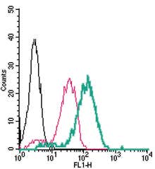 A Rabbit Polyclonal Antibody to CX3CR1 Conjugated to the Fluorescent Dye FITC
- Product Code: ACR-059-F
- Short Description: A Rabbit Polyclonal Antibody to CX3CR1 Conjugated to the Fluorescent Dye FITC
- Host: Rabbit
- Label: FITC
- Reactivity: Human
- Applications: Live cell imaging, Flow cytometry
- Target: Fractalkine Receptor, CX3C chemokine receptor 1, C-X3-C CKR-1, C-X3-C motif chemokine receptor 1, GPR13, CMKBRL1, GPRV28, V28, G-protein coupled receptor 13, CCRL1, Beta chemokine receptor-like 1
-
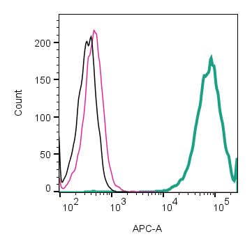 A rabbit polyclonal antibody to HVCN1 (extracellular)
- Product Code: AHC-002
- Short Description: A rabbit polyclonal antibody to HVCN1 (extracellular)
- Host: Rabbit
- Label: Unconjugated
- Reactivity: Rat, Mouse
- Applications: Indirect flow cytometry, Live cell imaging, Western blot
- Target: HVCN1
-
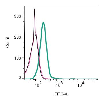
 A Rabbit Polyclonal Antibody to HVCN1 Channel
- Product Code: AHC-001
- Short Description: A Rabbit Polyclonal Antibody to HVCN1 Channel
- Host: Rabbit
- Label: Unconjugated
- Reactivity: Human, Rat, Mouse
- Applications: Indirect flow cytometry, Western blot
- Target: Hydrogen voltage-gated channel 1, HV1, Voltage sensor domain-only protein, VSOP
-
 A Rabbit Polyclonal Antibody to IBA1/AIF1
- Product Code: ACS-010
- Short Description: A Rabbit Polyclonal Antibody to IBA1/AIF1
- Host: Rabbit
- Label: Unconjugated
- Reactivity: Rat, Mouse
- Applications: Western blot, Immunofluorescence, Immunohistochemistry
- Target: Ionized calcium-binding adapter molecule 1, Allograft Inflammatory Factor 1, MRF-1, Microglia response factor
-
 A Rabbit Polyclonal Antibody to KCNK13 Channel
- Product Code: APC-121
- Short Description: A Rabbit Polyclonal Antibody to KCNK13 Channel
- Host: Rabbit
- Label: Unconjugated
- Reactivity: Rat, Mouse
- Applications: Indirect flow cytometry, Live cell imaging, Western blot, Immunohistochemistry
- Target: Potassium channel subfamily K member 13, Tandem pore domain halothane-inhibited K+ channel 1, K2P13.1
-
 A Rabbit Polyclonal Antibody to MERTK Receptor
- Product Code: ATR-033
- Short Description: A Rabbit Polyclonal Antibody to MERTK Receptor
- Host: Rabbit
- Label: Unconjugated
- Reactivity: Human, Rat, Mouse
- Applications: Indirect flow cytometry, Western blot, Immunofluorescence, Immunohistochemistry
- Target: Receptor tyrosine kinase MerTK, C-Mer Proto-Oncogene Tyrosine Kinase, Mer, Proto-Oncogene C-Mer
-
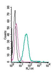 A Rabbit Polyclonal Antibody to MERTK (extracellular) conjugated to the fluorescent dye FITC
- Product Code: ATR-033-F
- Short Description: A Rabbit Polyclonal Antibody to MERTK (extracellular) conjugated to the fluorescent dye FITC
- Host: Rabbit
- Label: FITC
- Reactivity: Human, Rat, Mouse
- Applications: Live cell imaging, Flow cytometry
- Target: Receptor tyrosine kinase MerTK, C-Mer Proto-Oncogene Tyrosine Kinase, Mer, Proto-Oncogene C-Mer
-
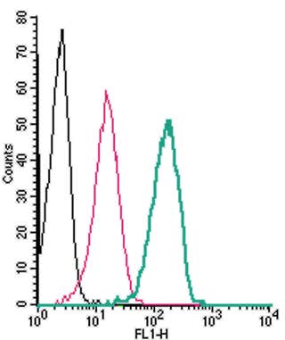 A Rabbit Polyclonal Antibody to P2X4 Receptor
- Product Code: APR-024
- Short Description: A Rabbit Polyclonal Antibody to P2X4 Receptor
- Host: Rabbit
- Label: Unconjugated
- Reactivity: Human, Rat, Mouse
- Applications: Indirect flow cytometry, Live cell imaging, Western blot, Immunocytochemistry, Immunofluorescence, Immunohistochemistry
- Target: P2RX4, P2X purinoceptor 4, ATP receptor
Loading...