-

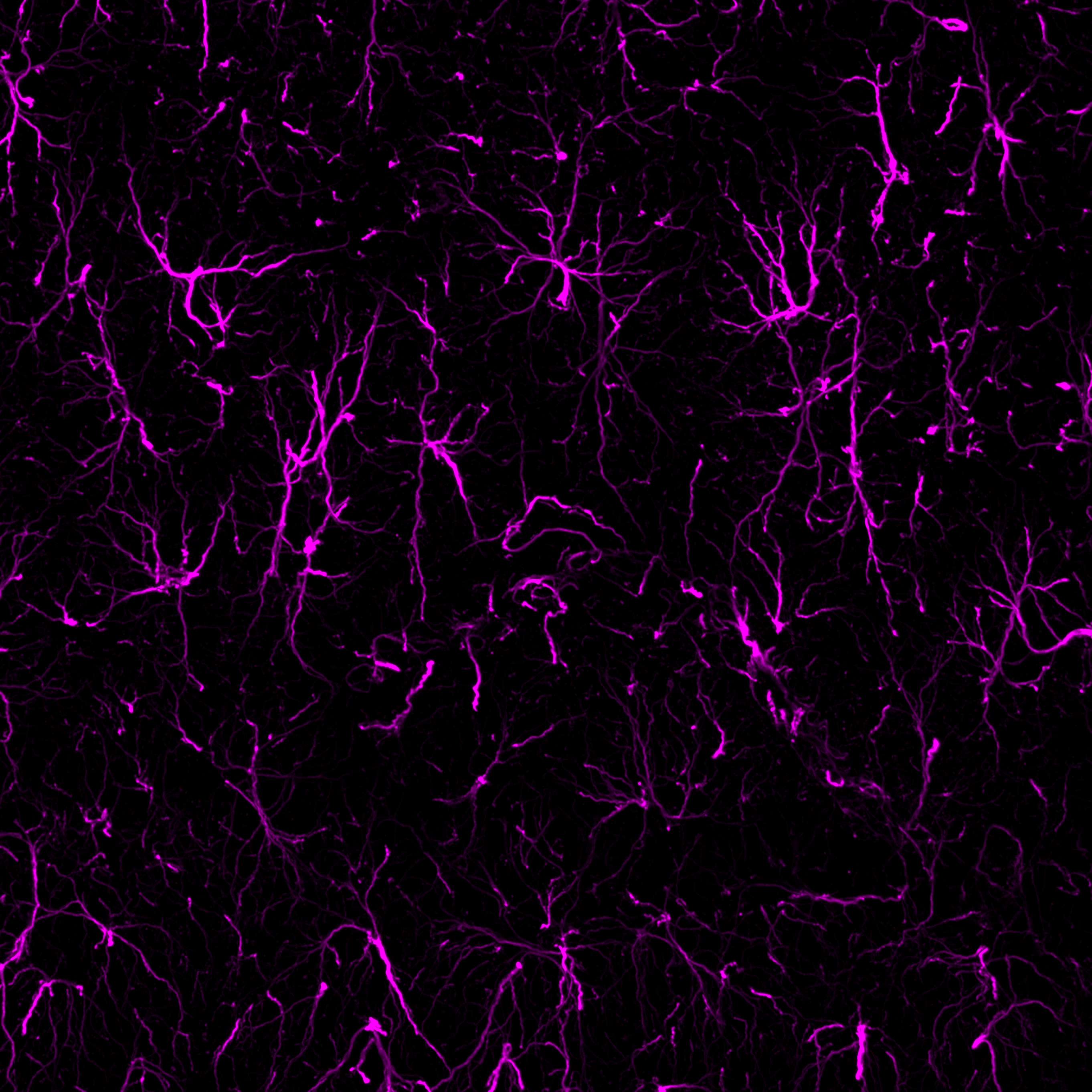
 A Rabbit Polyclonal Antibody to P2X7 Receptor
- Product Code: APR-004
- Short Description: A Rabbit Polyclonal Antibody to P2X7 Receptor
- Host: Rabbit
- Label: Unconjugated
- Reactivity: Human, Rat, Mouse
- Applications: Western blot, Immunocytochemistry, Immunofluorescence, Immunoprecipitation, Immunohistochemistry
- Target: Purinergic receptor P2X7, P2RX7, P2Z, ATP receptor
-
 A Rabbit Polyclonal Antibody to P2X7 Receptor Conjugated to the Fluorescent Dye ATTO-550
- Product Code: APR-004-AO
- Short Description: A Rabbit Polyclonal Antibody to P2X7 Receptor Conjugated to the Fluorescent Dye ATTO-550
- Host: Rabbit
- Label: ATTO 550
- Reactivity: Human, Rat, Mouse
- Applications: Immunofluorescence, Immunohistochemistry
- Target: Purinergic receptor P2X7, P2RX7, P2Z, ATP receptor
-
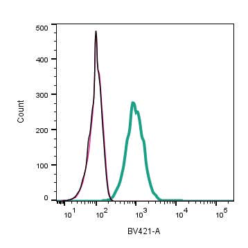 A Rabbit Polyclonal Antibody to P2Y12 Receptor
- Product Code: APR-020
- Short Description: A Rabbit Polyclonal Antibody to P2Y12 Receptor
- Host: Rabbit
- Label: Unconjugated
- Reactivity: Human, Mouse
- Applications: Indirect flow cytometry, Live cell imaging, Western blot, Immunocytochemistry, Immunofluorescence
- Target: P2Y purinoceptor 12, P2RY12, ADP-glucose receptor, Platelet ADP receptor, HORK3
-
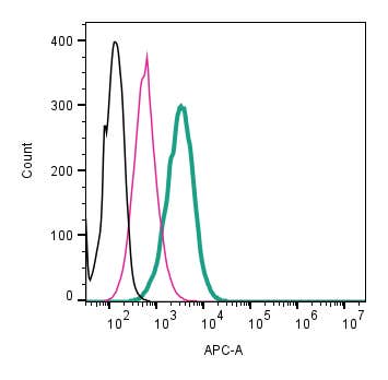 A Rabbit Polyclonal Antibody to P2Y12 Receptor (extracellular) conjugated to the fluorescent dye Allophycocyanin (APC).
- Product Code: APR-020-APC
- Short Description: A Rabbit Polyclonal Antibody to P2Y12 Receptor (extracellular) conjugated to the fluorescent dye Allophycocyanin (APC).
- Host: Rabbit
- Label: APC
- Reactivity: Human, Rat, Mouse
- Applications: Live cell imaging, Flow cytometry
- Target: P2Y purinoceptor 12, P2RY12, P2Y receptor
-
 A Rabbit Polyclonal Antibody to P2Y12 Receptor Conjugated to the Fuorescent dye FITC
- Product Code: APR-020-F
- Short Description: A Rabbit Polyclonal Antibody to P2Y12 Receptor Conjugated to the Fuorescent dye FITC
- Host: Rabbit
- Label: FITC
- Reactivity: Human, Rat, Mouse
- Applications: Live cell imaging, Flow cytometry
- Target: P2Y purinoceptor 12, P2RY12, ADP-glucose receptor, Platelet ADP receptor, HORK3
-
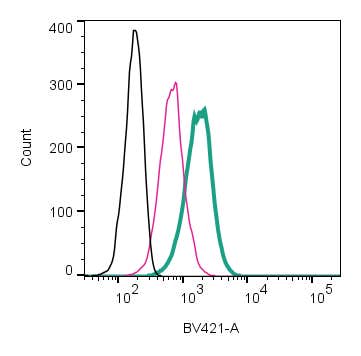 A Rabbit Polyclonal Antibody to P2Y12 Receptor conjugated to mFluor™ Violet 450 fluorophore
- Product Code: APR-020-V
- Short Description: A Rabbit Polyclonal Antibody to P2Y12 Receptor conjugated to mFluor™ Violet 450 fluorophore
- Host: Rabbit
- Label: mFluor™ Violet 450
- Reactivity: Human, Rat, Mouse
- Applications: Live cell imaging, Flow cytometry
- Target: P2Y purinoceptor 12, P2RY12, P2Y receptor
-
 A Rabbit Polyclonal Antibody to P2Y12 Conjugated to the Fluorescent Dye Phycoerythrin
- Product Code: APR-020-PE
- Short Description: A Rabbit Polyclonal Antibody to P2Y12 Conjugated to the Fluorescent Dye Phycoerythrin
- Host: Rabbit
- Label: R-PE
- Reactivity: Human, Rat, Mouse
- Applications: Live cell imaging, Flow cytometry
- Target: P2Y purinoceptor 12, P2RY12, P2Y receptor
-
 A Rabbit Polyclonal Antibody to P2Y12 Receptor
- Product Code: APR-012
- Short Description: A Rabbit Polyclonal Antibody to P2Y12 Receptor
- Host: Rabbit
- Label: Unconjugated
- Reactivity: Human, Rat, Mouse
- Applications: Indirect flow cytometry, Western blot, Immunocytochemistry, Immunofluorescence, Immunoprecipitation, Immunohistochemistry
- Target: P2Y purinoceptor 12, P2RY12, ADP-glucose receptor, Platelet ADP receptor, HORK3
-
 A Rabbit Polyclonal Antibody to P2Y12 Receptor Conjugated to the Fluorescent Dye ATTO-594
- Product Code: APR-012-AR
- Short Description: A Rabbit Polyclonal Antibody to P2Y12 Receptor Conjugated to the Fluorescent Dye ATTO-594
- Host: Rabbit
- Label: ATTO 594
- Reactivity: Human, Rat, Mouse
- Applications: Immunofluorescence, Immunohistochemistry
- Target: P2Y purinoceptor 12, P2RY12, ADP-glucose receptor, Platelet ADP receptor, HORK3
-
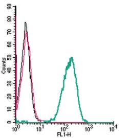 A Rabbit Polyclonal antibody to P2Y13 (extracellular).
- Product Code: APR-103
- Short Description: A Rabbit Polyclonal antibody to P2Y13 (extracellular).
- Host: Rabbit
- Label: Unconjugated
- Reactivity: Human, Rat, Mouse
- Applications: Indirect flow cytometry, Live cell imaging, Western blot
- Target: Purinergic Receptor P2Y13, P2RY13, GPCR1, GPR86, GPR94
Loading...