-
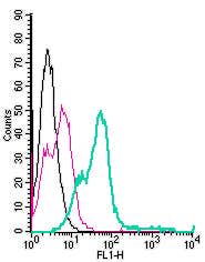 A Rabbit Polyclonal Antibody to Nogo Receptor Conjugated to the Fluorescent Dye FITC
- Product Code: ANT-008-F
- Short Description: A Rabbit Polyclonal Antibody to Nogo Receptor Conjugated to the Fluorescent Dye FITC
- Host: Rabbit
- Label: FITC
- Reactivity: Human, Rat, Mouse
- Applications: Flow cytometry
- Target: NogoR, Nogo-R, NgR, NgR1, Nogo-66 receptor, Reticulon 4 receptor, RTN4R
-
 A Rabbit Polyclonal Antibody to NPY1R (extracellular) conjugated to the fluorescent dye FITC
- Product Code: ANR-027-F
- Short Description: A Rabbit Polyclonal Antibody to NPY1R (extracellular) conjugated to the fluorescent dye FITC
- Host: Rabbit
- Label: FITC
- Reactivity: Human, Rat, Mouse
- Applications: Live cell imaging, Flow cytometry
- Target: Neuropeptide Y receptor type 1, NPYR, NPYY1
-
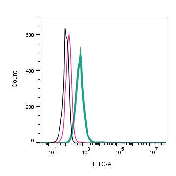 A Rabbit Polyclonal Antibody to NrCAM (extracellular) conjugated to the fluorescent dye FITC
- Product Code: ANR-045-F
- Short Description: A Rabbit Polyclonal Antibody to NrCAM (extracellular) conjugated to the fluorescent dye FITC
- Host: Rabbit
- Label: FITC
- Reactivity: Human, Rat, Mouse
- Applications: Live cell imaging, Flow cytometry
- Target: Neuronal cell adhesion molecule, Nr-CAM, NgCAM-related cell adhesion molecule, Ankyrin-binding cell adhesion molecule, Neuronal surface protein Bravo, HBravo
-
 A Rabbit Polyclonal Antibody to Neuregulin-1 Conjugated to the Fluorescent Dye ATTO-488
- Product Code: ANR-111-AG
- Short Description: A Rabbit Polyclonal Antibody to Neuregulin-1 Conjugated to the Fluorescent Dye ATTO-488
- Host: Rabbit
- Label: ATTO 488
- Reactivity: Mouse
- Applications: Immunofluorescence, Immunohistochemistry
- Target: Glial growth factor, GGF, Heregulin, HRG, Sensory and motor neuron derived factor, SMDF, Neu differentiation factor, NDF
-
 A Rabbit Polyclonal Antibody to Orai1 Channel Conjugated to the Fluorescent Dye ATTO-488
- Product Code: ACC-062-AG
- Short Description: A Rabbit Polyclonal Antibody to Orai1 Channel Conjugated to the Fluorescent Dye ATTO-488
- Host: Rabbit
- Label: ATTO 488
- Reactivity: Rat, Mouse
- Applications: Immunocytochemistry, Immunofluorescence
- Target: TMEM142A, CRACM1, Calcium release-activated calcium channel protein 1
-
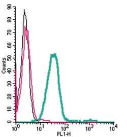 A Rabbit Polyclonal Antibody to Orai1 (extracellular) conjugated to the fluorescent dye FITC.
- Product Code: ACC-062-F
- Short Description: A Rabbit Polyclonal Antibody to Orai1 (extracellular) conjugated to the fluorescent dye FITC.
- Host: Rabbit
- Label: FITC
- Reactivity: Rat, Mouse
- Applications: Live cell imaging, Flow cytometry
- Target: TMEM142A, Calcium release-activated calcium channel protein 1, CRACM1
-
 A Rabbit Polyclonal Antibody to Orexin Receptor 1 Conjugated to the Fluorescent Dye ATTO-488
- Product Code: AOR-001-AG
- Short Description: A Rabbit Polyclonal Antibody to Orexin Receptor 1 Conjugated to the Fluorescent Dye ATTO-488
- Host: Rabbit
- Label: ATTO 488
- Reactivity: Human, Rat, Mouse
- Applications: Immunofluorescence, Immunohistochemistry
- Target: OX1R, Hypocretin receptor type 1, HCRTR1
-
 A Rabbit Polyclonal Antibody to P2X1 Receptor Conjugated to the Fluorescent Dye ATTO-488
- Product Code: APR-022-AG
- Short Description: A Rabbit Polyclonal Antibody to P2X1 Receptor Conjugated to the Fluorescent Dye ATTO-488
- Host: Rabbit
- Label: ATTO 488
- Reactivity: Human, Rat, Mouse
- Applications: Live cell imaging, Immunocytochemistry, Immunofluorescence, Immunohistochemistry
- Target: P2RX1, P2X purinoceptor 1
-
 A Rabbit Polyclonal Antibody to P2X1 Receptor Conjugated to the Fluorescent Dye FITC
- Product Code: APR-022-F
- Short Description: A Rabbit Polyclonal Antibody to P2X1 Receptor Conjugated to the Fluorescent Dye FITC
- Host: Rabbit
- Label: FITC
- Reactivity: Human, Rat, Mouse
- Applications: Live cell imaging, Flow cytometry
- Target: P2RX1, P2X purinoceptor 1
-

 A Rabbit Polyclonal Antibody to P2X2 Receptor Conjugated to the Fluorescent Dye ATTO-594
- Product Code: APR-003-AR
- Short Description: A Rabbit Polyclonal Antibody to P2X2 Receptor Conjugated to the Fluorescent Dye ATTO-594
- Host: Rabbit
- Label: ATTO 594
- Reactivity: Human, Rat, Mouse
- Applications: Immunofluorescence, Immunohistochemistry
- Target: P2X purinoceptor 2, P2RX2
Loading...