-
 A Rabbit Polyclonal Antibody to P2Y12 Receptor Conjugated to the Fluorescent Dye ATTO-594
- Product Code: APR-012-AR
- Short Description: A Rabbit Polyclonal Antibody to P2Y12 Receptor Conjugated to the Fluorescent Dye ATTO-594
- Host: Rabbit
- Label: ATTO 594
- Reactivity: Human, Rat, Mouse
- Applications: Immunofluorescence, Immunohistochemistry
- Target: P2Y purinoceptor 12, P2RY12, ADP-glucose receptor, Platelet ADP receptor, HORK3
-
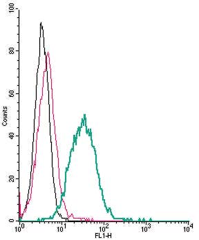 A Rabbit Polyclonal Antibody to P2Y13 Receptor (extracellular) conjugated to the fluorescent dye FITC
- Product Code: APR-103-F
- Short Description: A Rabbit Polyclonal Antibody to P2Y13 Receptor (extracellular) conjugated to the fluorescent dye FITC
- Host: Rabbit
- Label: FITC
- Reactivity: Human, Rat, Mouse
- Applications: Live cell imaging, Flow cytometry
- Target: Purinergic Receptor P2Y13, P2RY13, GPCR1, GPR86, GPR94, SP174
-
 A Rabbit Polyclonal Antibody to P2Y14 (extracellular) conjugated to the fluorescent dye FITC
- Product Code: APR-018-F
- Short Description: A Rabbit Polyclonal Antibody to P2Y14 (extracellular) conjugated to the fluorescent dye FITC
- Host: Rabbit
- Label: FITC
- Reactivity: Human, Rat, Mouse
- Applications: Live cell imaging, Flow cytometry
- Target: P2Y purinoceptor 14, P2RY14, GPR105, UDP-glucose receptor
-
 A Rabbit Polyclonal Antibody to P2Y2 Receptor Conjugated to the Fluorescent Dye FITC
- Product Code: APR-102-F
- Short Description: A Rabbit Polyclonal Antibody to P2Y2 Receptor Conjugated to the Fluorescent Dye FITC
- Host: Rabbit
- Label: FITC
- Reactivity: Human, Rat, Mouse
- Applications: Live cell imaging, Flow cytometry
- Target: P2Y purinoceptor 2, P2RY2, P2U1
-
 A Rabbit Polyclonal Antibody to P2Y6 Conjugated to the Fluorescent Dye FITC
- Product Code: APR-106-F
- Short Description: A Rabbit Polyclonal Antibody to P2Y6 Conjugated to the Fluorescent Dye FITC
- Host: Rabbit
- Label: FITC
- Reactivity: Human, Rat, Mouse
- Applications: Live cell imaging, Flow cytometry
- Target: P2Y purinoceptor 6, P2RY6
-
 A Rabbit Polyclonal Antibody to Human Neurotrophin Receptor p75NTR Conjugated to the Fluorescent Dye ATTO-488
- Product Code: ANT-007-AG
- Short Description: A Rabbit Polyclonal Antibody to Human Neurotrophin Receptor p75NTR Conjugated to the Fluorescent Dye ATTO-488
- Host: Rabbit
- Label: ATTO 488
- Reactivity: Human, Rat, Mouse
- Applications: Live cell imaging, Immunocytochemistry, Immunofluorescence, Flow cytometry, Immunohistochemistry
- Target: p75NTR, p75 Neurotrophin receptor, Low affinity NGF receptor, TNFR superfamily member 16, TNFRSF16, CD271
-
 A Rabbit Polyclonal Antibody to Human Neurotrophin Receptor p75NTR Conjugated to the Fluorescent Dye ATTO-550
- Product Code: ANT-007-AO
- Short Description: A Rabbit Polyclonal Antibody to Human Neurotrophin Receptor p75NTR Conjugated to the Fluorescent Dye ATTO-550
- Host: Rabbit
- Label: ATTO 550
- Reactivity: Human, Rat, Mouse
- Applications: Live cell imaging, Immunocytochemistry, Immunofluorescence, Immunohistochemistry
- Target: p75NTR, p75 Neurotrophin receptor, Low affinity NGF receptor, TNFR superfamily member 16, TNFRSF16, CD271
-
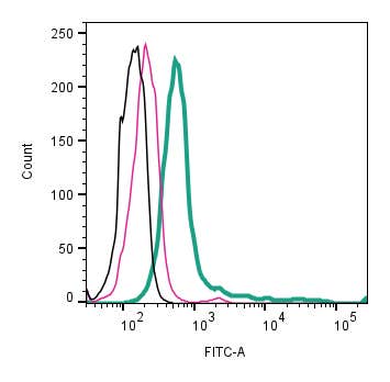 A Rabbit Polyclonal Antibody to Human Neurotrophin Receptor p75NTR Conjugated to the Fluorescent Dye FITC
- Product Code: ANT-007-F
- Short Description: A Rabbit Polyclonal Antibody to Human Neurotrophin Receptor p75NTR Conjugated to the Fluorescent Dye FITC
- Host: Rabbit
- Label: FITC
- Reactivity: Human, Rat, Mouse
- Applications: Live cell imaging, Flow cytometry
- Target: p75NTR, p75 Neurotrophin receptor, Low affinity NGF receptor, TNFR superfamily member 16, TNFRSF16, CD271
-

 A Rabbit Polyclonal Antibody to Protease-Activated Receptor-4 Conjugated to the Fluorescent Dye FITC
- Product Code: APR-034-F
- Short Description: A Rabbit Polyclonal Antibody to Protease-Activated Receptor-4 Conjugated to the Fluorescent Dye FITC
- Host: Rabbit
- Label: FITC
- Reactivity: Human, Rat, Mouse
- Applications: Live cell imaging, Flow cytometry
- Target: Protease-activated receptor-4, PAR-4, Coagulation factor II receptor-like 3, Thrombin receptor-like 3
-
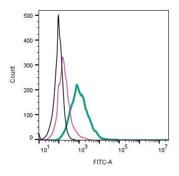 A Rabbit Polyclonal Antibody to Piezo1 (extracellular) conjugated to the fluorescent dye FITC
- Product Code: APC-087-F
- Short Description: A Rabbit Polyclonal Antibody to Piezo1 (extracellular) conjugated to the fluorescent dye FITC
- Host: Rabbit
- Label: FITC
- Reactivity: Rat, Mouse
- Applications: Live cell imaging, Flow cytometry
- Target: Piezo-type mechanosensitive ion channel component 1, Protein FAM38A, Mib, Membrane protein induced by beta-amyloid treatment
Loading...