-
 A Rabbit Polyclonal Antibody to Calcium Homeostasis Modulator 1 Conjugated to the Fluorescent Dye ATTO-594
- Product Code: ACC-101-AR
- Short Description: A Rabbit Polyclonal Antibody to Calcium Homeostasis Modulator 1 Conjugated to the Fluorescent Dye ATTO-594
- Host: Rabbit
- Label: ATTO 594
- Reactivity: Human, Rat, Mouse
- Applications: Immunocytochemistry, Immunofluorescence, Immunohistochemistry
- Target: Calcium homeostasis modulator 1, FAM26C
-
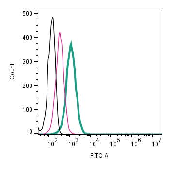 A Rabbit Polyclonal antibody to CALHM2 (extracellular) conjugated to the fluorescent dye FITC
- Product Code: ACC-142-F
- Short Description: A Rabbit Polyclonal antibody to CALHM2 (extracellular) conjugated to the fluorescent dye FITC
- Host: Rabbit
- Label: FITC
- Reactivity: Human, Rat, Mouse
- Applications: Live cell imaging, Flow cytometry
- Target: Calcium homeostasis modulator protein 2, Protein FAM26B, FAM26B
-
 A Rabbit Polyclonal Antibody to Calnexin Conjugated to the Fluorescent Dye ATTO-594
- Product Code: ACS-009-AR
- Short Description: A Rabbit Polyclonal Antibody to Calnexin Conjugated to the Fluorescent Dye ATTO-594
- Host: Rabbit
- Label: ATTO 594
- Reactivity: Human, Rat, Mouse
- Applications: Immunofluorescence, Immunohistochemistry
- Target: Canx, Calx, CNX, Major histocompatibility complex class I antigen-binding protein p88, p90, IP90
-
 A Rabbit Polyclonal Antibody to Cannabinoid Receptor 1 Conjugated to the Fluorescent Dye FITC
- Product Code: ACR-001-F
- Short Description: A Rabbit Polyclonal Antibody to Cannabinoid Receptor 1 Conjugated to the Fluorescent Dye FITC
- Host: Rabbit
- Label: FITC
- Reactivity: Human, Rat, Mouse
- Applications: Live cell imaging, Flow cytometry
- Target: CB1, CB-R, CNR1, CB1R, CANN6, Brain-type cannabinoid receptor, Central cannabinoid receptor
-

 A Rabbit Polyclonal Antibody to CaV1.2 (CACNA1C) Channel Conjugated to the Fluorescent Dye ATTO-488
- Product Code: ACC-003-AG
- Short Description: A Rabbit Polyclonal Antibody to CaV1.2 (CACNA1C) Channel Conjugated to the Fluorescent Dye ATTO-488
- Host: Rabbit
- Label: ATTO 488
- Reactivity: Human, Rat, Mouse
- Applications: Immunocytochemistry, Immunofluorescence, Immunohistochemistry
- Target: Voltage-dependent L-type calcium channel subunit α1C
-
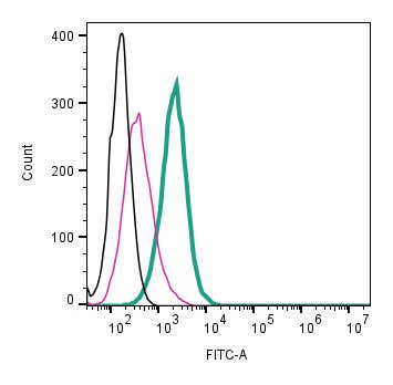 A Rabbit Polyclonal Antibody to CaV1.3 (CACNA1D) (extracellular) conjugated to the fluorescent dye FITC
- Product Code: ACC-311-F
- Short Description: A Rabbit Polyclonal Antibody to CaV1.3 (CACNA1D) (extracellular) conjugated to the fluorescent dye FITC
- Host: Rabbit
- Label: FITC
- Reactivity: Human, Rat, Mouse
- Applications: Live cell imaging, Flow cytometry
- Target: Voltage-dependent L-type calcium channel subunit α1D
-
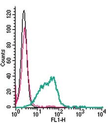 A Rabbit Polyclonal Antibody to CD39 Conjugated to the Fluorescent Dye FITC
- Product Code: ANT-065-F
- Short Description: A Rabbit Polyclonal Antibody to CD39 Conjugated to the Fluorescent Dye FITC
- Host: Rabbit
- Label: FITC
- Reactivity: Human, Rat, Mouse
- Applications: Live cell imaging, Flow cytometry
- Target: Ectonucleoside Triphosphate Diphosphohydrolase 1, Ecto-ATP diphosphohydrolase 1, Lymphoid cell activation antigen, ENTPD1
-
 A Rabbit Polyclonal Antibody to CD56/NCAM1
- Product Code: ANR-041-AG
- Short Description: A Rabbit Polyclonal Antibody to CD56/NCAM1
- Host: Rabbit
- Label: ATTO 488
- Reactivity: Human, Rat, Mouse
- Applications: Immunohistochemistry
- Target: NCAM-1, CD56
-
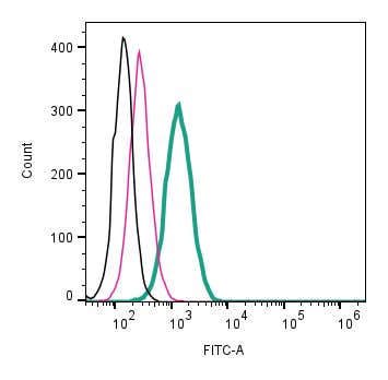 A Rabbit Polyclonal antibody to CD63 (extracellular) conjugated to the fluorescent dye FITC
- Product Code: ANR-210-F
- Short Description: A Rabbit Polyclonal antibody to CD63 (extracellular) conjugated to the fluorescent dye FITC
- Host: Rabbit
- Label: FITC
- Reactivity: Rat, Mouse
- Applications: Live cell imaging, Flow cytometry
- Target: Tetraspanin-30, TSPAN30, Lysosomal-Associated Membrane Protein 3, LAMP-3, Granulophysin, Melanoma-Associated Antigen ME491
-
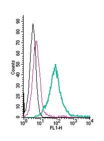 A Rabbit Polyclonal Antibody to CD73 (extracellular) conjugated to the fluorescent dye FITC
- Product Code: ANT-066-F
- Short Description: A Rabbit Polyclonal Antibody to CD73 (extracellular) conjugated to the fluorescent dye FITC
- Host: Rabbit
- Label: FITC
- Reactivity: Human, Rat, Mouse
- Applications: Flow cytometry
- Target: Ecto-5-nucleotidase, E5NT, 5'-Nucleotidase, 5'-NT, NT5E, ENT
Loading...