-
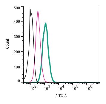 A Rabbit Polyclonal Antibody to CD81 (extracellular) (extracellular) conjugated to the fluorescent dye FITC
- Product Code: ANR-211-F
- Short Description: A Rabbit Polyclonal Antibody to CD81 (extracellular) (extracellular) conjugated to the fluorescent dye FITC
- Host: Rabbit
- Label: FITC
- Reactivity: Human, Rat, Mouse
- Applications: Live cell imaging, Flow cytometry
- Target: Tetraspanin-28, TSPAN28, Target of the Antiproliferative Antibody 1, TAPA-1
-
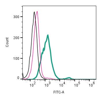 A Rabbit Polyclonal antibody to CD9 (extracellular) conjugated to the fluorescent dye FITC
- Product Code: ANR-209-F
- Short Description: A Rabbit Polyclonal antibody to CD9 (extracellular) conjugated to the fluorescent dye FITC
- Host: Rabbit
- Label: FITC
- Reactivity: Human, Rat, Mouse
- Applications: Live cell imaging, Flow cytometry
- Target: Tetrapanin-29, TSPAN29, Leukocyte Antigen MIC3, MIC3
-
 A Rabbit Polyclonal Antibody to CHERP Conjugated to the Fluorescent Dye ATTO-594
- Product Code: ACC-330-AR
- Short Description: A Rabbit Polyclonal Antibody to CHERP Conjugated to the Fluorescent Dye ATTO-594
- Host: Rabbit
- Label: ATTO 594
- Reactivity: Human, Rat, Mouse
- Applications: Immunofluorescence, Immunohistochemistry
- Target: Calcium homeostasis endoplasmic reticulum protein, SR-related CTD-associated factor 6, SCAF6
-
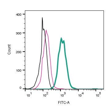 A Rabbit Polyclonal Antibody to CLEC7A (extracellular) conjugated to the fluorescent dye FITC
- Product Code: ALR-062-F
- Short Description: A Rabbit Polyclonal Antibody to CLEC7A (extracellular) conjugated to the fluorescent dye FITC
- Host: Rabbit
- Label: FITC
- Reactivity: Rat, Mouse
- Applications: Live cell imaging, Flow cytometry
- Target: C-Type Lectin Domain Family 7 Member A, Dendritic Cell-Associated C-Type Lectin 1, DC-Associated C-Type Lectin 1, Beta-Glucan Receptor, C-Type Lectin Superfamily Member 12, CLECSF12, CD369, SCARE2
-
 A Rabbit Polyclonal Antibody to Connexin-36 Conjugated to the Fluorescent Dye ATTO-488
- Product Code: ACC-209-AG
- Short Description: A Rabbit Polyclonal Antibody to Connexin-36 Conjugated to the Fluorescent Dye ATTO-488
- Host: Rabbit
- Label: ATTO 488
- Reactivity: Mouse
- Applications: Immunofluorescence, Immunohistochemistry
- Target: CX36, Gap junction δ2 protein, Gap junction α9 protein, GJD2, GJA9
-
 A Rabbit Polyclonal Antibody to Corticotropin-Releasing Factor Receptor 1 Conjugated to the Fluorescent Dye FITC
- Product Code: ACR-050-F
- Short Description: A Rabbit Polyclonal Antibody to Corticotropin-Releasing Factor Receptor 1 Conjugated to the Fluorescent Dye FITC
- Host: Rabbit
- Label: FITC
- Reactivity: Human, Rat, Mouse
- Applications: Live cell imaging, Flow cytometry
- Target: Corticotropin-releasing factor receptor 1, CRFR1, Corticotropin-releasing hormone receptor 1, CRH-R1, CRH1
-
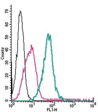 A Rabbit Polyclonal Antibody to CRLR/CALCRL (extracellular) conjugated to the fluorescent dye FITC
- Product Code: ACR-060-F
- Short Description: A Rabbit Polyclonal Antibody to CRLR/CALCRL (extracellular) conjugated to the fluorescent dye FITC
- Host: Rabbit
- Label: FITC
- Reactivity: Human, Rat, Mouse
- Applications: Live cell imaging, Flow cytometry
- Target: Calcitonin Receptor-Like Receptor, Calcitonin Gene-Related Peptide Type 1 Receptor, CRL, CGRPR
-
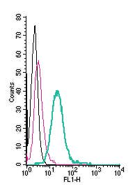 A Rabbit Polyclonal Antibody to CX3CR1 (extracellular) conjugated to the fluorescent dye FITC
- Product Code: ACR-058-F
- Short Description: A Rabbit Polyclonal Antibody to CX3CR1 (extracellular) conjugated to the fluorescent dye FITC
- Host: Rabbit
- Label: FITC
- Reactivity: Rat, Mouse
- Applications: Live cell imaging, Flow cytometry
- Target: Fractalkine Receptor, CX3C chemokine receptor 1, C-X3-C CKR-1, C-X3-C motif chemokine receptor 1, GPR13, CMKBRL1, GPRV28, V28, G-protein coupled receptor 13, CCRL1, Beta chemokine receptor-like 1
-
 A Rabbit Polyclonal Antibody to CXCR4 Chemokine Receptor Conjugated to the Fluorescent Dye ATTO-488
- Product Code: ACR-014-AG
- Short Description: A Rabbit Polyclonal Antibody to CXCR4 Chemokine Receptor Conjugated to the Fluorescent Dye ATTO-488
- Host: Rabbit
- Label: ATTO 488
- Reactivity: Human, Rat, Mouse
- Applications: Live cell imaging, Flow cytometry
- Target: C-X-C chemokine receptor type 4, CD184, Fusin, LESTR, NPYRL, SDF-1 receptor, WHIMS
-
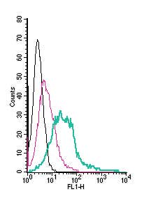 A Rabbit Polyclonal Antibody to CXCR4 (extracellular) conjugated to the fluorescent dye FITC
- Product Code: ACR-014-F
- Short Description: A Rabbit Polyclonal Antibody to CXCR4 (extracellular) conjugated to the fluorescent dye FITC
- Host: Rabbit
- Label: FITC
- Reactivity: Human, Rat, Mouse
- Applications: Live cell imaging, Flow cytometry
- Target: C-X-C chemokine receptor type 4, CD184, Fusin, LESTR, NPYRL, SDF-1 receptor, WHIMS
Loading...