-
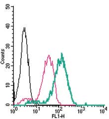 A Rabbit Polyclonal Antibody to CX3CR1 Conjugated to the Fluorescent Dye FITC
- Product Code: ACR-059-F
- Short Description: A Rabbit Polyclonal Antibody to CX3CR1 Conjugated to the Fluorescent Dye FITC
- Host: Rabbit
- Label: FITC
- Reactivity: Human
- Applications: Live cell imaging, Flow cytometry
- Target: Fractalkine Receptor, CX3C chemokine receptor 1, C-X3-C CKR-1, C-X3-C motif chemokine receptor 1, GPR13, CMKBRL1, GPRV28, V28, G-protein coupled receptor 13, CCRL1, Beta chemokine receptor-like 1
-
 A Rabbit Polyclonal Antibody to CXCR2 Chemokine Receptor Conjugated to the Fluorescent Dye ATTO-488
- Product Code: ACR-012-AG
- Short Description: A Rabbit Polyclonal Antibody to CXCR2 Chemokine Receptor Conjugated to the Fluorescent Dye ATTO-488
- Host: Rabbit
- Label: ATTO 488
- Reactivity: Human
- Applications: Live cell imaging, Flow cytometry
- Target: C-X-C chemokine receptor type 2, Interleukin-8 receptor B, IL-8R B, IL8RB, IL-8 receptor type 2, CD182
-
 A Rabbit Polyclonal Antibody to FPR1 Conjugated to the Fluorescent Dye ATTO-488
- Product Code: AFR-001-AG
- Short Description: A Rabbit Polyclonal Antibody to FPR1 Conjugated to the Fluorescent Dye ATTO-488
- Host: Rabbit
- Label: ATTO 488
- Reactivity: Human
- Applications: Live cell imaging, Flow cytometry
- Target: N-formyl peptide receptor 1, fMLP receptor
-
 A Rabbit Polyclonal Antibody to GLUT3 Conjugated to the Fluorescent Dye FITC
- Product Code: AGT-043-F
- Short Description: A Rabbit Polyclonal Antibody to GLUT3 Conjugated to the Fluorescent Dye FITC
- Host: Rabbit
- Label: FITC
- Reactivity: Human
- Applications: Flow cytometry
- Target: Glucose transporter 3, Facilitated glucose transporter member 3, SLC2A3
-
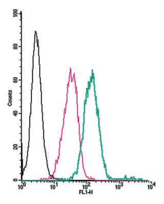 A Rabbit Polyclonal Antibody to Human GPR132 (extracellular) conjugated to the fluorescent dye FITC
- Product Code: AGR-046-F
- Short Description: A Rabbit Polyclonal Antibody to Human GPR132 (extracellular) conjugated to the fluorescent dye FITC
- Host: Rabbit
- Label: FITC
- Reactivity: Human
- Applications: Live cell imaging, Flow cytometry
- Target: G protein-coupled receptor 132, G2 accumulation protein, G2A
-
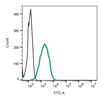 A Rabbit Polyclonal Antibody to Human Orai1 Channel Conjugated to the Fluorescent Dye FITC
- Product Code: ACC-060-F
- Short Description: A Rabbit Polyclonal Antibody to Human Orai1 Channel Conjugated to the Fluorescent Dye FITC
- Host: Rabbit
- Label: FITC
- Reactivity: Human
- Applications: Flow cytometry
- Target: TMEM142A, CRACM1, Calcium release-activated calcium channel protein 1
-
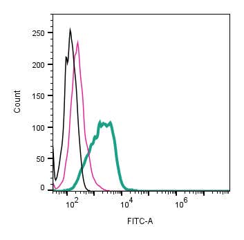 A Rabbit Polyclonal Antibody to Protease-Activated Receptor-1 Conjugated to the Fluorescent dye FITC
- Product Code: APR-031-F
- Short Description: A Rabbit Polyclonal Antibody to Protease-Activated Receptor-1 Conjugated to the Fluorescent dye FITC
- Host: Rabbit
- Label: FITC
- Reactivity: Human
- Applications: Live cell imaging, Flow cytometry
- Target: Protease-activated receptor-1, PAR-1, Thrombin receptor, Coagulation factor II receptor, CF2R
-
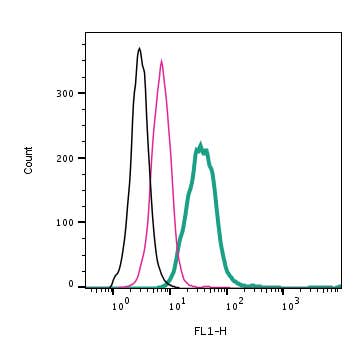 A Rabbit Polyclonal Antibody to PAR2 (extracellular) conjugated to the fluorescent dye FITC
- Product Code: APR-035-F
- Short Description: A Rabbit Polyclonal Antibody to PAR2 (extracellular) conjugated to the fluorescent dye FITC
- Host: Rabbit
- Label: FITC
- Reactivity: Human
- Applications: Live cell imaging, Flow cytometry
- Target: Protease-activated receptor-2, PAR-2, Thrombin receptor-like 1, Coagulation factor II receptor-like 1, GPR11
-
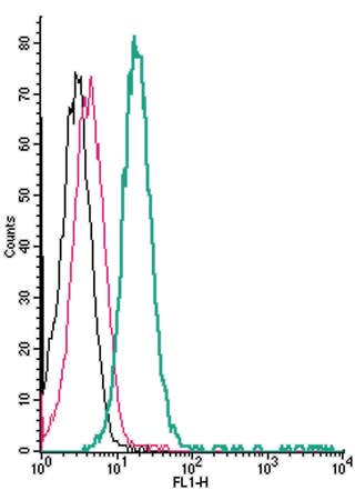 A Rabbit Polyclonal Antibody Human TRPV1 (extracellular) conjugated to the fluorescent dye FITC
- Product Code: ACC-334-F
- Short Description: A Rabbit Polyclonal Antibody Human TRPV1 (extracellular) conjugated to the fluorescent dye FITC
- Host: Rabbit
- Label: FITC
- Reactivity: Human
- Applications: Live cell imaging, Flow cytometry
- Target: Transient receptor potential cation channel subfamily V member 1, Capsaicin receptor, Vanilloid receptor 1, VR1, OTRPC1
-
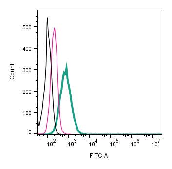 A Rabbit Polyclonal Antibody to Human TRPV2 (extracellular) conjugated to the fluorescent dye FITC
- Product Code: ACC-139-F
- Short Description: A Rabbit Polyclonal Antibody to Human TRPV2 (extracellular) conjugated to the fluorescent dye FITC
- Host: Rabbit
- Label: FITC
- Reactivity: Human
- Applications: Live cell imaging, Flow cytometry
- Target: Vanilloid receptor-like protein 1, VRL-1, Osm-9-like TRP channel 2, OTRPC2
Loading...