-
 A Rabbit Polyclonal Antibody to STIM1 Conjugated to the Fluorescent Dye ATTO-550
- Product Code: ACC-063-AO
- Short Description: A Rabbit Polyclonal Antibody to STIM1 Conjugated to the Fluorescent Dye ATTO-550
- Host: Rabbit
- Label: ATTO 550
- Reactivity: Human, Rat, Mouse
- Applications: Live cell imaging, Immunocytochemistry, Immunofluorescence, Flow cytometry, Immunohistochemistry
- Target: Stromal interaction molecule 1, GOK
-
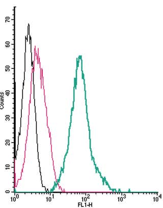 A Rabbit Polyclonal Antibody to Thromboxane A2 Receptor (extracellular) conjugated to the fluorescent dye FITC
- Product Code: APR-095-F
- Short Description: A Rabbit Polyclonal Antibody to Thromboxane A2 Receptor (extracellular) conjugated to the fluorescent dye FITC
- Host: Rabbit
- Label: FITC
- Reactivity: Human, Rat, Mouse
- Applications: Live cell imaging, Flow cytometry
- Target: Prostanoid TP receptor, TXA2-R, TBXA2R
-
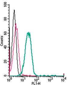 A Rabbit Polyclonal Antibody to TMEM119 (extracellular) conjugated to the fluorescent dye FITC
- Product Code: ANR-175-F
- Short Description: A Rabbit Polyclonal Antibody to TMEM119 (extracellular) conjugated to the fluorescent dye FITC
- Host: Rabbit
- Label: FITC
- Reactivity: Rat, Mouse
- Applications: Flow cytometry
- Target: Transmembrane Protein 119, Osteoblast induction factor, OBIF
-
 A Rabbit Polyclonal Antibody to TMEM66 Conjugated to the Fluorescent Dye ATTO-594
- Product Code: ACC-067-AR
- Short Description: A Rabbit Polyclonal Antibody to TMEM66 Conjugated to the Fluorescent Dye ATTO-594
- Host: Rabbit
- Label: ATTO 594
- Reactivity: Human, Rat, Mouse
- Applications: Immunofluorescence, Immunohistochemistry
- Target: Store-operated calcium entry-associated regulatory factor, SOCE-associated regulatory factor, SARAF, Transmembrane protein 66
-
 A Rabbit Polyclonal Antibody to TREM2 Conjugated to the Fluorescent Dye FITC
- Product Code: ANR-018-F
- Short Description: A Rabbit Polyclonal Antibody to TREM2 Conjugated to the Fluorescent Dye FITC
- Host: Rabbit
- Label: FITC
- Reactivity: Human, Rat, Mouse
- Applications: Live cell imaging, Flow cytometry
- Target: Triggering receptor expressed on myeloid cells 2, Triggering receptor expressed on monocytes 2
-
 A Rabbit Polyclonal Antibody to TrkA Conjugated to the Fluorescent Dye ATTO-633
- Product Code: ANT-018-FR
- Short Description: A Rabbit Polyclonal Antibody to TrkA Conjugated to the Fluorescent Dye ATTO-633
- Host: Rabbit
- Label: ATTO 633
- Reactivity: Human, Rat, Mouse
- Applications: Immunofluorescence, Immunohistochemistry
- Target: High affinity nerve growth factor receptor, Neurotrophic tyrosine kinase receptor type 1, NTRK1
-
 A Rabbit Polyclonal Antibody to TrkA Conjugated to the Fluorescent Dye FITC
- Product Code: ANT-018-F
- Short Description: A Rabbit Polyclonal Antibody to TrkA Conjugated to the Fluorescent Dye FITC
- Host: Rabbit
- Label: FITC
- Reactivity: Human, Rat, Mouse
- Applications: Flow cytometry
- Target: High affinity nerve growth factor receptor, Neurotrophic tyrosine kinase receptor type 1, NTRK1
-
 A Rabbit Polyclonal Antibody to TrkB Conjugated to the Fluorescent Dye ATTO-488
- Product Code: ANT-019-AG
- Short Description: A Rabbit Polyclonal Antibody to TrkB Conjugated to the Fluorescent Dye ATTO-488
- Host: Rabbit
- Label: ATTO 488
- Reactivity: Human, Rat, Mouse
- Applications: Immunofluorescence, Flow cytometry, Immunohistochemistry
- Target: Neurotrophic tyrosine kinase receptor type 2, NTRK2, BDNF/NT-3 growth factors receptor
-
 A Rabbit Polyclonal Antibody to TrkB Conjugated to the fluorescent dye FITC
- Product Code: ANT-019-F
- Short Description: A Rabbit Polyclonal Antibody to TrkB Conjugated to the fluorescent dye FITC
- Host: Rabbit
- Label: FITC
- Reactivity: Human, Rat, Mouse
- Applications: Live cell imaging, Flow cytometry
- Target: Neurotrophic tyrosine kinase receptor type 2, NTRK2, BDNF/NT-3 growth factors receptor
-
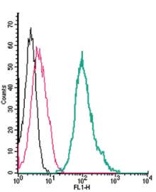 A Rabbit Polyclonal Antibody to TRPA1 (extracellular) conjugated to the fluorescent dye FITC
- Product Code: ACC-037-F
- Short Description: A Rabbit Polyclonal Antibody to TRPA1 (extracellular) conjugated to the fluorescent dye FITC
- Host: Rabbit
- Label: FITC
- Reactivity: Human, Rat, Mouse
- Applications: Flow cytometry
- Target: Transient receptor potential cation channel subfamily A member 1, ANKTM1, Ankyrin-like with transmembrane domains protein 1, Transformation-sensitive protein p120, Wasabi Receptor
Loading...