-
 A Rabbit Polyclonal Antibody to Glucose Transporter 4 Conjugated to the Fluorescent Dye ATTO-594
- Product Code: AGT-024-AR
- Short Description: A Rabbit Polyclonal Antibody to Glucose Transporter 4 Conjugated to the Fluorescent Dye ATTO-594
- Host: Rabbit
- Label: ATTO 594
- Reactivity: Human, Rat, Mouse
- Applications: Immunofluorescence, Immunohistochemistry
- Target: Glucose transporter 4, Facilitated glucose transporter member 4, Insulin-responsive glucose transporter type 4, SLC2A4
-
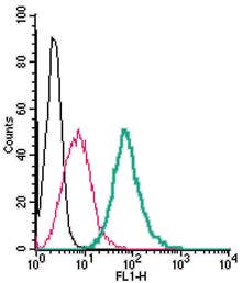 A Rabbit Polyclonal Antibody to GPER1 Conjugated to the Fluorescent Dye FITC
- Product Code: AER-049-F
- Short Description: A Rabbit Polyclonal Antibody to GPER1 Conjugated to the Fluorescent Dye FITC
- Host: Rabbit
- Label: FITC
- Reactivity: Human, Rat, Mouse
- Applications: Flow cytometry
- Target: G Protein-Coupled Estrogen Receptor 1, G Protein-Coupled Receptor 30, Membrane Estrogen Receptor, Chemoattractant Receptor-Like 2, GPER
-
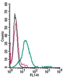 A Rabbit Polyclonal Antibody to GPR120/FFAR4 Conjugated to the Fluorescent Dye FITC
- Product Code: AFR-014-F
- Short Description: A Rabbit Polyclonal Antibody to GPR120/FFAR4 Conjugated to the Fluorescent Dye FITC
- Host: Rabbit
- Label: FITC
- Reactivity: Human, Rat, Mouse
- Applications: Flow cytometry
- Target: G-protein coupled receptor 120, Free fatty acid receptor 4, FFA4 Receptor, G-protein coupled receptor GT01, Omega-3 fatty acid receptor 1, O3far1
-
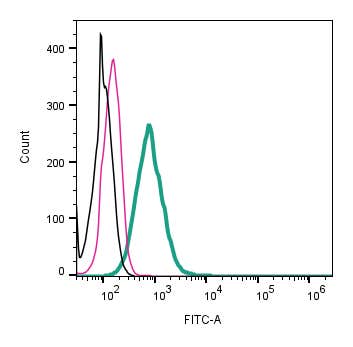 A Rabbit Polyclonal Antibody to GPR142 (extracellular) conjugated to the fluorescent dye FITC
- Product Code: AGR-082-F
- Short Description: A Rabbit Polyclonal Antibody to GPR142 (extracellular) conjugated to the fluorescent dye FITC
- Host: Rabbit
- Label: FITC
- Reactivity: Human, Rat, Mouse
- Applications: Live cell imaging, Flow cytometry
- Target: G-Protein Coupled Receptor 142, Probable G-Protein Coupled Receptor 142, PGR2
-
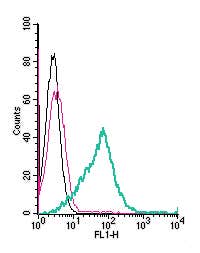 A Rabbit Polyclonal Antibody to GPR174 (extracellular) conjugated to the fluorescent dye FITC
- Product Code: AGR-060-F
- Short Description: A Rabbit Polyclonal Antibody to GPR174 (extracellular) conjugated to the fluorescent dye FITC
- Host: Rabbit
- Label: FITC
- Reactivity: Human, Rat, Mouse
- Applications: Live cell imaging, Flow cytometry
- Target: G-protein coupled receptor 174, LYPSR3, GPCR17, FKSG79
-
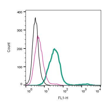 A Rabbit Polyclonal Antibody to GPR183 (extracellular) conjugated to the fluorescent dye FITC
- Product Code: AGR-063-F
- Short Description: A Rabbit Polyclonal Antibody to GPR183 (extracellular) conjugated to the fluorescent dye FITC
- Host: Rabbit
- Label: FITC
- Reactivity: Human, Rat, Mouse
- Applications: Live cell imaging, Flow cytometry
- Target: G-Protein Coupled Receptor 183, Epstein-Barr Virus-Induced G-Protein Coupled Receptor 2, EBI2
-
 A Rabbit Polyclonal Antibody to GPR34 Conjugated to the Fluorescent Dye FITC
- Product Code: AGR-055-F
- Short Description: A Rabbit Polyclonal Antibody to GPR34 Conjugated to the Fluorescent Dye FITC
- Host: Rabbit
- Label: FITC
- Reactivity: Human, Rat, Mouse
- Applications: Flow cytometry
- Target: G-protein coupled receptor 34
-
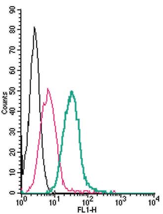 A Rabbit Polyclonal Antibody to GPR35 (extracellular) conjugated to the fluorescent dye FITC
- Product Code: AGR-051-F
- Short Description: A Rabbit Polyclonal Antibody to GPR35 (extracellular) conjugated to the fluorescent dye FITC
- Host: Rabbit
- Label: FITC
- Reactivity: Rat, Mouse
- Applications: Live cell imaging, Flow cytometry
- Target: G-Protein Coupled Receptor 35, Kynurenic Acid Receptor
-
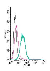 A Rabbit Polyclonal Antibody to GPR4 (extracellular) conjugated to the fluorescent dye FITC
- Product Code: AGR-041-F
- Short Description: A Rabbit Polyclonal Antibody to GPR4 (extracellular) conjugated to the fluorescent dye FITC
- Host: Rabbit
- Label: FITC
- Reactivity: Human, Rat, Mouse
- Applications: Live cell imaging, Flow cytometry
- Target: Acid-sensing G-protein coupled receptor 4, GPR19
-
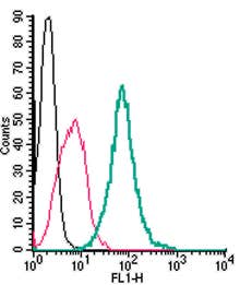 A Rabbit Polyclonal Antibody to GPR55 Conjugated to the Fluorescent Dye FITC
- Product Code: ACR-062-F
- Short Description: A Rabbit Polyclonal Antibody to GPR55 Conjugated to the Fluorescent Dye FITC
- Host: Rabbit
- Label: FITC
- Reactivity: Rat, Mouse
- Applications: Live cell imaging, Flow cytometry
- Target: G-Protein Coupled Receptor 55
Loading...