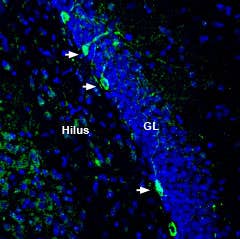
-
 A Rabbit Polyclonal Antibody to NMDA Receptor 2A (GluN2A) Conjugated to the Fluorescent Dye ATTO-488
- Product Code: AGC-002-AG
- Short Description: A Rabbit Polyclonal Antibody to NMDA Receptor 2A (GluN2A) Conjugated to the Fluorescent Dye ATTO-488
- Host: Rabbit
- Label: ATTO 488
- Reactivity: Human, Rat, Mouse
- Applications: Immunofluorescence, Immunohistochemistry
- Target: NMDA receptor 2A, NR2A, GRIN2A, Ionotropic glutamate receptor subunit ε1, N-methyl-D-aspartate receptor subunit 2A
-
 A Rabbit Polyclonal Antibody to NMDA Receptor 2A (GluN2A)
- Product Code: AGC-002
- Short Description: A Rabbit Polyclonal Antibody to NMDA Receptor 2A (GluN2A)
- Host: Rabbit
- Label: Unconjugated
- Reactivity: Human, Rat, Mouse
- Applications: Indirect flow cytometry, Live cell imaging, Western blot, Immunocytochemistry, Immunofluorescence, Immunoprecipitation, Immunohistochemistry
- Target: NMDA receptor 2A, NR2A, GRIN2A, Ionotropic glutamate receptor subunit ε1, N-methyl-D-aspartate receptor subunit 2A
-
 A Guinea Pig Polyclonal Antibody to NMDA Receptor 1 (GluN1)
- Product Code: AGC-001-GP
- Short Description: A Guinea Pig Polyclonal Antibody to NMDA Receptor 1 (GluN1)
- Host: Guinea Pig
- Label: Unconjugated
- Reactivity: Human, Rat, Mouse
- Applications: Western blot, Immunofluorescence, Immunohistochemistry
- Target: NMDA receptor 1, Ionotropic glutamate receptor subunit ζ 1, N-methyl-D-aspartate receptor subunit 1, GRIN1
-
 A Rabbit Polyclonal Antibody to NMDA Receptor 1 (GluN1)
- Product Code: AGC-001
- Short Description: A Rabbit Polyclonal Antibody to NMDA Receptor 1 (GluN1)
- Host: Rabbit
- Label: Unconjugated
- Reactivity: Human, Rat, Mouse
- Applications: Indirect flow cytometry, Live cell imaging, Western blot, Immunocytochemistry, Immunofluorescence, Immunohistochemistry
- Target: NMDA receptor 1, Ionotropic glutamate receptor subunit ζ 1, N-methyl-D-aspartate receptor subunit 1, GRIN1
-
 A Rabbit Polyclonal Antibody to GABA(B) Receptor 2
- Product Code: AGB-002
- Short Description: A Rabbit Polyclonal Antibody to GABA(B) Receptor 2
- Host: Rabbit
- Label: Unconjugated
- Reactivity: Human, Rat, Mouse
- Applications: Western blot, Immunofluorescence, Immunohistochemistry
- Target: γ-Aminobutyric acid type B receptor subunit 2, GABA B receptor 2, GABABR2, Gb2, GABBR2, GPR51, GPRC3B, HG20
-
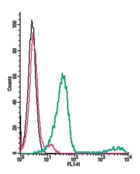 A Rabbit Polyclonal Antibody to GABBR1 (extracellular) conjugated to the fluorescent dye FITC
- Product Code: AGB-001-F
- Short Description: A Rabbit Polyclonal Antibody to GABBR1 (extracellular) conjugated to the fluorescent dye FITC
- Host: Rabbit
- Label: FITC
- Reactivity: Human, Rat, Mouse
- Applications: Live cell imaging, Flow cytometry
- Target: γ-Aminobutyric acid type B receptor subunit 1, GABA B receptor 1, GABABR1, Gb1, GABBR1, GPRC3A
-
 A Rabbit Polyclonal Antibody to GABA(B) Receptor 1
- Product Code: AGB-001
- Short Description: A Rabbit Polyclonal Antibody to GABA(B) Receptor 1
- Host: Rabbit
- Label: Unconjugated
- Reactivity: Human, Rat, Mouse
- Applications: Indirect flow cytometry, Live cell imaging, Western blot, Immunofluorescence, Immunohistochemistry
- Target: γ-Aminobutyric acid type B receptor subunit 1, GABA B receptor 1, GABABR1, Gb1, GABBR1, GPRC3A
-

 A Rabbit Polyclonal Antibody to GABA(A) δ Receptor
- Product Code: AGA-014
- Short Description: A Rabbit Polyclonal Antibody to GABA(A) δ Receptor
- Host: Rabbit
- Label: Unconjugated
- Reactivity: Rat, Mouse
- Applications: Indirect flow cytometry, Live cell imaging, Western blot, Immunofluorescence, Immunohistochemistry
- Target: GABRD, Gamma-aminobutyric acid receptor subunit delta, GABA(A) receptor subunit delta
-
 A Rabbit Polyclonal Antibody to GABA(A) β1 Receptor
- Product Code: AGA-010
- Short Description: A Rabbit Polyclonal Antibody to GABA(A) β1 Receptor
- Host: Rabbit
- Label: Unconjugated
- Reactivity: Human, Rat, Mouse
- Applications: Western blot
- Target: γ-Aminobutyric acid receptor subunit β1, GABAA receptor beta-1, GABRB1, GARB1
-
 A Rabbit Polyclonal Antibody to GABA(A) α4 Receptor
- Product Code: AGA-008
- Short Description: A Rabbit Polyclonal Antibody to GABA(A) α4 Receptor
- Host: Rabbit
- Label: Unconjugated
- Reactivity: Human, Rat, Mouse
- Applications: Live cell imaging, Western blot, Immunocytochemistry, Immunofluorescence, Immunohistochemistry
- Target: γ-Aminobutyric acid receptor subunit α4, GABRA4
Loading...