-
 A Rabbit Polyclonal Antibody to Calnexin Conjugated to the Fluorescent Dye ATTO-594
- Product Code: ACS-009-AR
- Short Description: A Rabbit Polyclonal Antibody to Calnexin Conjugated to the Fluorescent Dye ATTO-594
- Host: Rabbit
- Label: ATTO 594
- Reactivity: Human, Rat, Mouse
- Applications: Immunofluorescence, Immunohistochemistry
- Target: Canx, Calx, CNX, Major histocompatibility complex class I antigen-binding protein p88, p90, IP90
-
 A Rabbit Polyclonal Antibody to CaV1.1 (CACNA1S) Channel
- Product Code: ACC-314
- Short Description: A Rabbit Polyclonal Antibody to CaV1.1 (CACNA1S) Channel
- Host: Rabbit
- Label: Unconjugated
- Reactivity: Human, Rat, Mouse
- Applications: Western blot, Immunohistochemistry
- Target: Voltage-dependent L-type calcium channel subunit α1S, CACH1, CACN1, CACH1B, CACNL1A3
-
 A Rabbit Polyclonal Antibody to CaV1.3 (CACNA1D) Channel
- Product Code: ACC-311
- Short Description: A Rabbit Polyclonal Antibody to CaV1.3 (CACNA1D) Channel
- Host: Rabbit
- Label: Unconjugated
- Reactivity: Human, Rat, Mouse
- Applications: Indirect flow cytometry, Live cell imaging, Western blot, Immunocytochemistry, Immunofluorescence, Immunohistochemistry
- Target: Voltage-dependent L-type calcium channel subunit α1D
-
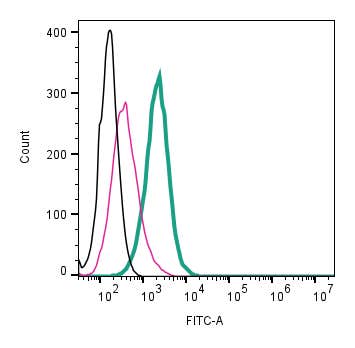 A Rabbit Polyclonal Antibody to CaV1.3 (CACNA1D) (extracellular) conjugated to the fluorescent dye FITC
- Product Code: ACC-311-F
- Short Description: A Rabbit Polyclonal Antibody to CaV1.3 (CACNA1D) (extracellular) conjugated to the fluorescent dye FITC
- Host: Rabbit
- Label: FITC
- Reactivity: Human, Rat, Mouse
- Applications: Live cell imaging, Flow cytometry
- Target: Voltage-dependent L-type calcium channel subunit α1D
-

 A Rabbit Polyclonal Antibody to CaV1.3 (CACNA1D) Channel
- Product Code: ACC-005
- Short Description: A Rabbit Polyclonal Antibody to CaV1.3 (CACNA1D) Channel
- Host: Rabbit
- Label: Unconjugated
- Reactivity: Human, Rat, Mouse
- Applications: Western blot, Immunocytochemistry, Immunofluorescence, Immunoprecipitation, Immunohistochemistry
- Target: Voltage-dependent L-type calcium channel subunit α1D
-
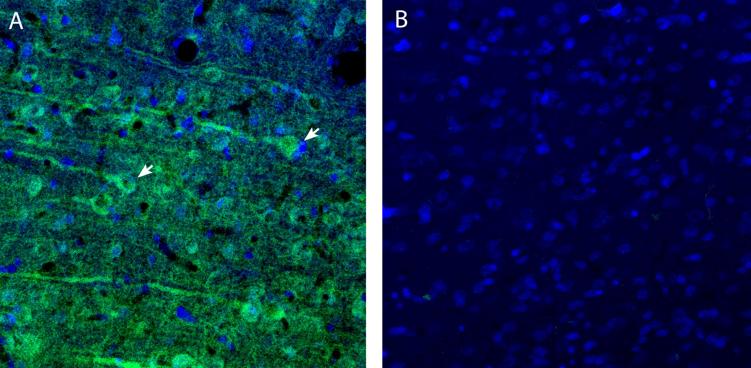 A rabbit polyclonal antibody to CCC9 (SLC12A8) (extracellular)
- Product Code: AKT-009
- Short Description: A rabbit polyclonal antibody to CCC9 (SLC12A8) (extracellular)
- Host: Rabbit
- Label: Unconjugated
- Reactivity: Human, Rat, Mouse
- Applications: Western blot, Immunohistochemistry
- Target: SLC12A8
-
 A Rabbit Polyclonal Antibody to CCR7 Chemokine Receptor
- Product Code: ACR-027
- Short Description: A Rabbit Polyclonal Antibody to CCR7 Chemokine Receptor
- Host: Rabbit
- Label: Unconjugated
- Reactivity: Human, Rat
- Applications: Indirect flow cytometry, Live cell imaging, Western blot, Immunohistochemistry
- Target: C-C chemokine receptor type 7, C-C CKR-7, Epstein-Barr virus-induced G-protein coupled receptor 1, EBI1, CD197
-
 A Rabbit Polyclonal antibody to C3aR1 (extracellular)
- Product Code: ANT-067
- Short Description: A Rabbit Polyclonal antibody to C3aR1 (extracellular)
- Host: Rabbit
- Label: Unconjugated
- Reactivity: Rat, Mouse
- Applications: Indirect flow cytometry, Live cell imaging, Western blot, Immunohistochemistry
- Target: Bone Marrow Stromal Antigen 1, BST-1, ADP-Ribosyl Cyclase 2, Cyclic ADP-ribose hydrolase 2, CADPr Hydrolase 2, CADPR2, LY-65
-
 A Rabbit Polyclonal antibody to CD38 (extracellular)
- Product Code: ANT-068
- Short Description: A Rabbit Polyclonal antibody to CD38 (extracellular)
- Host: Rabbit
- Label: Unconjugated
- Reactivity: Rat, Mouse
- Applications: Indirect flow cytometry, Live cell imaging, Western blot, Immunohistochemistry
- Target: ADP-Ribosyl Cyclase 1, ADPRC 1, Cyclic ADP-Ribose Hydrolase 1, cADPR hydrolase 1, CADPR1
-
 A Rabbit Polyclonal Antibody to CD39
- Product Code: ANT-065
- Short Description: A Rabbit Polyclonal Antibody to CD39
- Host: Rabbit
- Label: Unconjugated
- Reactivity: Human, Rat
- Applications: Indirect flow cytometry, Live cell imaging, Western blot
- Target: Ectonucleoside triphosphate diphosphohydrolase 1, NTPDase 1, Ecto-ATP diphosphohydrolase 1, Ecto-ATPDase 1, Ecto-apyrase, Lymphoid cell activation antigen, ENTPD1
Loading...