-

 A potent antagonist of muscle nAChRs, α7 nAChRs, and GABA(A) receptor subtypes
- Product Code: B-100
- Short Description: A potent antagonist of muscle nAChRs, α7 nAChRs, and GABA(A) receptor subtypes
- Applications: Electrophysiology
- CAS No.: 11032-79-4
- MW: 7984 Da
-
 A fluorescent probe for imaging and sensitive detection of neuromuscular junctions, GABA(A) receptors, nAChRs, and α-Bungarotoxin binding sites
- Product Code: B-100-AG
- Short Description: A fluorescent probe for imaging and sensitive detection of neuromuscular junctions, GABA(A) receptors, nAChRs, and α-Bungarotoxin binding sites
- Label: ATTO 488
- Applications: Electrophysiology, Direct flow cytometry, Fluorescence staining
- MW: ~9140 Da
-
 A fluorescent probe for imaging and sensitive detection of neuromuscular junctions, GABA(A) receptors, nAChRs, and α-Bungarotoxin binding sites
- Product Code: B-100-FR
- Short Description: A fluorescent probe for imaging and sensitive detection of neuromuscular junctions, GABA(A) receptors, nAChRs, and α-Bungarotoxin binding sites
- Label: ATTO 633
- Applications: Electrophysiology, Direct flow cytometry, Fluorescence staining, Live cell imaging, Immunofluorescence
- MW: ~9140 Da
-
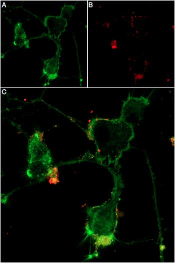 A fluorescent probe for imaging and sensitive detection of neuromuscular junctions, GABA(A) receptors, nAChRs, and α-Bungarotoxin binding sites
- Product Code: B-100-FRN
- Short Description: A fluorescent probe for imaging and sensitive detection of neuromuscular junctions, GABA(A) receptors, nAChRs, and α-Bungarotoxin binding sites
- Label: ATTO 647N
- Applications: Electrophysiology, Direct flow cytometry, Fluorescence staining, Live cell imaging, Immunofluorescence
- MW: ~8925 Da
-
 A biotinylated probe for imaging and sensitive detection of neuromuscular junctions, GABA(A) receptors, nAChRs, and α-Bungarotoxin binding sites
- Product Code: B-100-B
- Short Description: A biotinylated probe for imaging and sensitive detection of neuromuscular junctions, GABA(A) receptors, nAChRs, and α-Bungarotoxin binding sites
- Label: Biotin
- Applications: Electrophysiology, Immunofluorescence, Indirect flow cytometry
- MW: ~9030 Da
-
 A fluorescent probe for imaging and sensitive detection of neuromuscular junctions, GABA(A) receptors, nAChRs, and α-Bungarotoxin binding sites
- Product Code: B-100-F
- Short Description: A fluorescent probe for imaging and sensitive detection of neuromuscular junctions, GABA(A) receptors, nAChRs, and α-Bungarotoxin binding sites
- Label: FITC
- Applications: Electrophysiology, Fluorescence staining
- MW: ~8406 Da
-
 A potent antagonist of the neuronal α3β2 and α7 nicotinic acetylcholine receptors (nAChR)
- Product Code: C-290
- Short Description: A potent antagonist of the neuronal α3β2 and α7 nicotinic acetylcholine receptors (nAChR)
- Applications: Electrophysiology
- CAS No.: 156467-85-5
- MW: 1351 Da
-
 An α7 nAChR potent antagonist that functions as a PAM-dependent biased agonist
- Product Code: STC-320
- Short Description: An α7 nAChR potent antagonist that functions as a PAM-dependent biased agonist
- Applications: Electrophysiology
- MW: 1835 Da
-
 A potent and selective antagonist of neuronal α6*-containing nicotinic acetylcholine receptors (nAChRs)
- Product Code: STC-870
- Short Description: A potent and selective antagonist of neuronal α6*-containing nicotinic acetylcholine receptors (nAChRs)
- Applications: Electrophysiology
- CAS No.: 669050-68-4
- MW: 1981 Da
-
 A potent antagonist of α9α10 nAChRs that also inhibits N-type (CaV2.2) calcium channels via GABA(B) receptor activation
- Product Code: STC-010
- Short Description: A potent antagonist of α9α10 nAChRs that also inhibits N-type (CaV2.2) calcium channels via GABA(B) receptor activation
- Applications: Electrophysiology
- MW: 1571 Da
Loading...