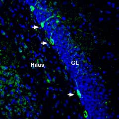
-

 A Rabbit Polyclonal Antibody to SCN1A (NaV1.1) Channel
- Product Code: ASC-001
- Short Description: A Rabbit Polyclonal Antibody to SCN1A (NaV1.1) Channel
- Host: Rabbit
- Label: Unconjugated
- Reactivity: Human, Rat, Mouse
- Applications: Western blot, Immunocytochemistry, Immunofluorescence, Immunoprecipitation, Immunohistochemistry
- Target: Brain type I sodium channel, BI, Sodium channel protein type 1 subunit α
-
 A Rabbit Polyclonal Antibody to PSD-95
- Product Code: APZ-009
- Short Description: A Rabbit Polyclonal Antibody to PSD-95
- Host: Rabbit
- Label: Unconjugated
- Reactivity: Human, Rat, Mouse
- Applications: Western blot, Immunofluorescence, Immunohistochemistry
- Target: Postsynaptic density protein 95, Discs large homolog 4, DLG4, Synapse-associated protein 90, SAP-90, Tax interaction protein 15
-
 A Rabbit Polyclonal Antibody to P2Y12 Conjugated to the Fluorescent Dye Phycoerythrin
- Product Code: APR-020-PE
- Short Description: A Rabbit Polyclonal Antibody to P2Y12 Conjugated to the Fluorescent Dye Phycoerythrin
- Host: Rabbit
- Label: R-PE
- Reactivity: Human, Rat, Mouse
- Applications: Live cell imaging, Flow cytometry
- Target: P2Y purinoceptor 12, P2RY12, P2Y receptor
-
 A Rabbit Polyclonal Antibody to P2Y12 Receptor Conjugated to the Fuorescent dye FITC
- Product Code: APR-020-F
- Short Description: A Rabbit Polyclonal Antibody to P2Y12 Receptor Conjugated to the Fuorescent dye FITC
- Host: Rabbit
- Label: FITC
- Reactivity: Human, Rat, Mouse
- Applications: Live cell imaging, Flow cytometry
- Target: P2Y purinoceptor 12, P2RY12, ADP-glucose receptor, Platelet ADP receptor, HORK3
-
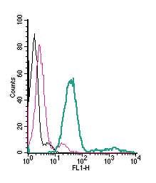 A Rabbit Polyclonal Antibody to P2X7 Receptor Conjugated to the Fluorescent Dye FITC
- Product Code: APR-008-F
- Short Description: A Rabbit Polyclonal Antibody to P2X7 Receptor Conjugated to the Fluorescent Dye FITC
- Host: Rabbit
- Label: FITC
- Reactivity: Human, Rat, Mouse
- Applications: Live cell imaging, Immunocytochemistry, Immunofluorescence, Flow cytometry
- Target: Purinergic receptor P2X7, P2RX7, P2Z, ATP receptor
-
 A Rabbit Polyclonal Antibody to P2X7 Receptor
- Product Code: APR-008
- Short Description: A Rabbit Polyclonal Antibody to P2X7 Receptor
- Host: Rabbit
- Label: Unconjugated
- Reactivity: Human, Rat, Mouse
- Applications: Indirect flow cytometry, Live cell imaging, Western blot, Immunocytochemistry, Immunofluorescence, Immunohistochemistry
- Target: Purinergic receptor P2X7, P2RX7, P2Z, ATP receptor
-
 A Rabbit Polyclonal Antibody to KV1.2 (KCNA2) Channel
- Product Code: APC-010
- Short Description: A Rabbit Polyclonal Antibody to KV1.2 (KCNA2) Channel
- Host: Rabbit
- Label: Unconjugated
- Reactivity: Human, Rat, Mouse
- Applications: Western blot, Immunocytochemistry, Immunofluorescence, Immunohistochemistry
- Target: Potassium voltage-gated channel subfamily A member 2, RBK2
-
 A Rabbit Polyclonal Antibody to TrkB
- Product Code: ANT-019
- Short Description: A Rabbit Polyclonal Antibody to TrkB
- Host: Rabbit
- Label: Unconjugated
- Reactivity: Human, Rat, Mouse
- Applications: Indirect flow cytometry, Live cell imaging, Western blot, Immunocytochemistry, Immunofluorescence, Immunohistochemistry
- Target: Neurotrophic tyrosine kinase receptor type 2, NTRK2, BDNF/NT-3 growth factors receptor
-
 A Rabbit Polyclonal Antibody to TrkA
- Product Code: ANT-018
- Short Description: A Rabbit Polyclonal Antibody to TrkA
- Host: Rabbit
- Label: Unconjugated
- Reactivity: Human, Rat, Mouse
- Applications: Indirect flow cytometry, Live cell imaging, Western blot, Immunocytochemistry, Immunofluorescence, Immunohistochemistry
- Target: High affinity nerve growth factor receptor, Neurotrophic tyrosine kinase receptor type 1, NTRK1
-
 A Rabbit Polyclonal Antibody to Human Neurotrophin Receptor p75NTR
- Product Code: ANT-007
- Short Description: A Rabbit Polyclonal Antibody to Human Neurotrophin Receptor p75NTR
- Host: Rabbit
- Label: Unconjugated
- Reactivity: Human, Rat, Mouse
- Applications: Indirect flow cytometry, Live cell imaging, Western blot, Immunocytochemistry, Immunofluorescence, Immunoprecipitation, Immunohistochemistry
- Target: p75NTR, p75 Neurotrophin receptor, Low affinity NGF receptor, TNFR superfamily member 16, TNFRSF16, CD271
Loading...