-
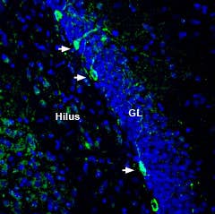
 A Rabbit Polyclonal Antibody to Nicotinic Acetylcholine Receptor α3
- Product Code: ANC-003
- Short Description: A Rabbit Polyclonal Antibody to Nicotinic Acetylcholine Receptor α3
- Host: Rabbit
- Label: Unconjugated
- Reactivity: Rat, Mouse
- Applications: Western blot, Immunofluorescence, Immunohistochemistry
- Target: nAChR α3, Neuronal acetylcholine receptor subunit alpha-3, ACHA3, PAOD2
-
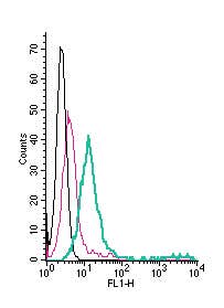 A Rabbit Polyclonal Antibody to GABRA1 (extracellular) conjugated to the fluorescent dye FITC
- Product Code: AGA-001-F
- Short Description: A Rabbit Polyclonal Antibody to GABRA1 (extracellular) conjugated to the fluorescent dye FITC
- Host: Rabbit
- Label: FITC
- Reactivity: Human, Rat, Mouse
- Applications: Live cell imaging, Flow cytometry
- Target: γ-Aminobutyric acid receptor type A subunit α1, GABRA1
-
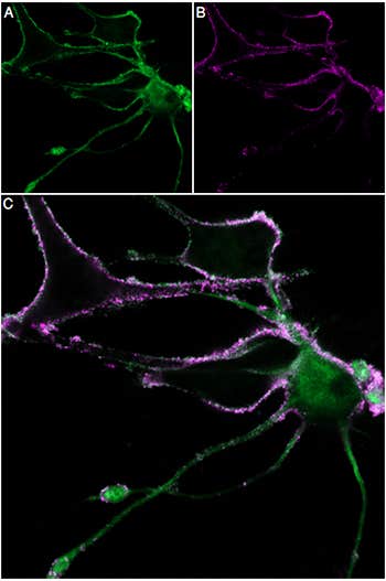
 A fluorescent probe for imaging and sensitive detection of Kv1.1, Kv1.2, and Kv1.6 channels.
- Product Code: D-350-FRN
- Short Description: A fluorescent probe for imaging and sensitive detection of Kv1.1, Kv1.2, and Kv1.6 channels.
- Label: ATTO 647N
- Applications: Electrophysiology, Direct flow cytometry, Fluorescence staining, Live cell imaging, Immunofluorescence
- MW: ~7990 Da
-
 A Blocking Peptide for Ant-OXGR1/GPR99 (extracellular) Antibody
- Product Code: BLP-OR017
- Short Description: A Blocking Peptide for Ant-OXGR1/GPR99 (extracellular) Antibody
- Applications: Western blot, Immunohistochemistry
- Target: GPR99
-
 A Blocking Peptide for Anti-CHRM3 (extracellular) Antibody
- Product Code: BLP-MR013
- Short Description: A Blocking Peptide for Anti-CHRM3 (extracellular) Antibody
- Applications: Western blot, Immunohistochemistry
- Target: CHRM3
-
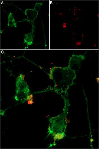 A fluorescent probe for imaging and sensitive detection of neuromuscular junctions, GABA(A) receptors, nAChRs, and α-Bungarotoxin binding sites
- Product Code: B-100-FRN
- Short Description: A fluorescent probe for imaging and sensitive detection of neuromuscular junctions, GABA(A) receptors, nAChRs, and α-Bungarotoxin binding sites
- Label: ATTO 647N
- Applications: Electrophysiology, Direct flow cytometry, Fluorescence staining, Live cell imaging, Immunofluorescence
- MW: ~8925 Da
-
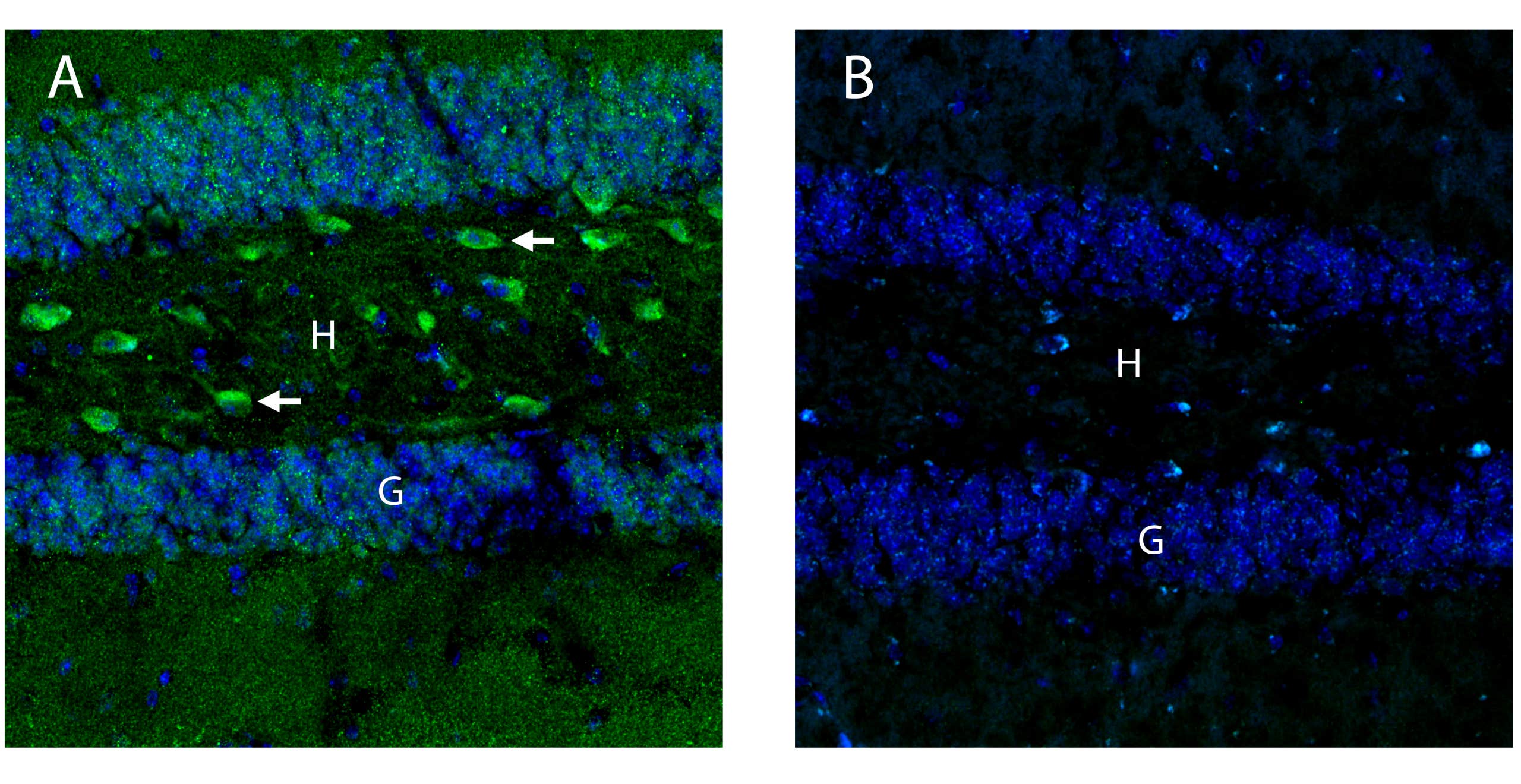 A rabbit polyclonal antibody to OXGR1/GPR99 (extracellular)
- Product Code: AOR-017
- Short Description: A rabbit polyclonal antibody to OXGR1/GPR99 (extracellular)
- Host: Rabbit
- Label: Unconjugated
- Reactivity: Human, Rat, Mouse
- Applications: Indirect flow cytometry, Western blot, Immunohistochemistry
- Target: Oxoglutarate receptor 1, 2-Oxoglutarate receptor 1, Alpha-ketoglutarate receptor 1, AKGR, GPR80, P2RY15, P2Y15, CysLTR3
-
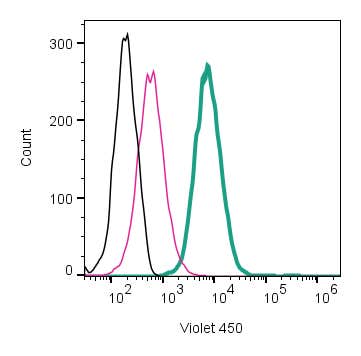 A Rabbit Polyclonal Antibody to C3aR1 (extracellular) conjugated to the fluorescent dye mFluor™ Violet 450 fluorophore.
- Product Code: AAR-036-V
- Short Description: A Rabbit Polyclonal Antibody to C3aR1 (extracellular) conjugated to the fluorescent dye mFluor™ Violet 450 fluorophore.
- Host: Rabbit
- Label: mFluor™ Violet 450
- Reactivity: Rat, Mouse
- Applications: Live cell imaging, Flow cytometry
- Target: Complement C3a Receptor 1, C3AR, C3a Anaphylatoxin Chemotactic Receptor
-
 A fluorescent probe for imaging and sensitive detection of KV1.2, KV1.3, KV1.1, and KV1.6 potassium channels
- Product Code: STT-360-AR
- Short Description: A fluorescent probe for imaging and sensitive detection of KV1.2, KV1.3, KV1.1, and KV1.6 potassium channels
- Label: ATTO 594
- Applications: Electrophysiology, Direct flow cytometry, Live cell imaging, Immunofluorescence
- MW: ~4900 Da
-
 A fluorescent probe for imaging and sensitive detection of Kir1.1 and GIRK potassium channels
- Product Code: STT-170-FR
- Short Description: A fluorescent probe for imaging and sensitive detection of Kir1.1 and GIRK potassium channels
- Label: ATTO 633
- Applications: Electrophysiology, Fluorescence staining, Live cell imaging, Immunofluorescence
- MW: 3159.6 Da
Loading...