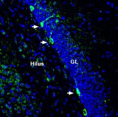
-

 A fluorescent probe for imaging and sensitive detection of Kir1.1 and GIRK potassium channels
- Product Code: STT-170-AG
- Short Description: A fluorescent probe for imaging and sensitive detection of Kir1.1 and GIRK potassium channels
- Label: ATTO 488
- Applications: Electrophysiology, Direct flow cytometry, Live cell imaging, Immunofluorescence
- MW: 3195.5 Da
-
 A biotinylated probe for imaging and sensitive detection of multiple KV1 channels (KV1.1, KV1.3, KV1.4, KV1.6) and KCa3.1 channels.
- Product Code: STS-400-B
- Short Description: A biotinylated probe for imaging and sensitive detection of multiple KV1 channels (KV1.1, KV1.3, KV1.4, KV1.6) and KCa3.1 channels.
- Label: Biotin
- Applications: Electrophysiology, Immunofluorescence, Indirect flow cytometry
- MW: 4563 Da
-
 A fluorescent probe for imaging and sensitive detection of multiple KV1 channels (KV1.1, KV1.3, KV1.4, KV1.6) and KCa3.1 channels.
- Product Code: STS-400-AR
- Short Description: A fluorescent probe for imaging and sensitive detection of multiple KV1 channels (KV1.1, KV1.3, KV1.4, KV1.6) and KCa3.1 channels.
- Label: ATTO 590
- Applications: Electrophysiology, Direct flow cytometry, Live cell imaging, Immunofluorescence
- MW: ~4627 Da
-
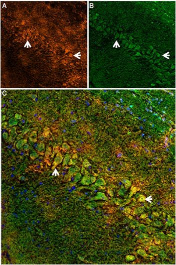 A fluorescent probe for imaging and sensitive detection of GABA(A) receptors
- Product Code: STM-550-AG
- Short Description: A fluorescent probe for imaging and sensitive detection of GABA(A) receptors
- Label: ATTO 488
- Applications: Electrophysiology, Direct flow cytometry, Live cell imaging, Immunofluorescence
- MW: ~7775 Da
-
 A biotinylated apamin for imaging and sensitive detection of KCa2 potassium channels (SK channels)
- Product Code: STA-200-B
- Short Description: A biotinylated apamin for imaging and sensitive detection of KCa2 potassium channels (SK channels)
- Label: Biotin
- Applications: Electrophysiology, Immunofluorescence
- MW: 2367 Da
-
 A fluorescent probe for imaging and sensitive detection of KV1.3, KV1.1, and KV1.6 voltage-gated potassium channels
- Product Code: RTA-420-T
- Short Description: A fluorescent probe for imaging and sensitive detection of KV1.3, KV1.1, and KV1.6 voltage-gated potassium channels
- Label: TAMRA
- Applications: Electrophysiology, Fluorescence staining
- MW: 4673 Da
-
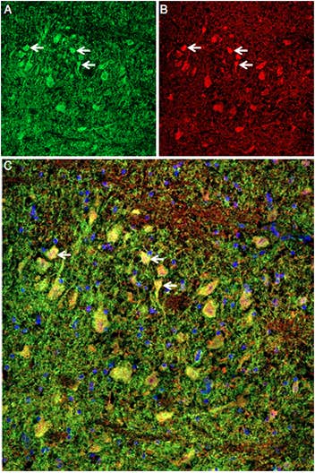 A fluorescent probe for imaging and sensitive detection of α3β2 and α7 nicotinic acetylcholine receptors (nAChR)
- Product Code: C-290-AR
- Short Description: A fluorescent probe for imaging and sensitive detection of α3β2 and α7 nicotinic acetylcholine receptors (nAChR)
- Label: ATTO 590
- Applications: Electrophysiology, Fluorescence staining, Live cell imaging, Immunofluorescence
- MW: 1924 Da
-
 A Blocking Peptide for Anti-ZIP8/SLC39A8 (extracellular) Antibody
- Product Code: BLP-ZT008
- Short Description: A Blocking Peptide for Anti-ZIP8/SLC39A8 (extracellular) Antibody
- Applications: Western blot, Immunohistochemistry
- Target: SLC39A8
-
 A Blocking Peptide for Anti-Human ZACN (extracellular) Antibody
- Product Code: BLP-ZC001
- Short Description: A Blocking Peptide for Anti-Human ZACN (extracellular) Antibody
- Applications: Western blot, Immunohistochemistry
- Target: ZACN
-
 A Blocking Peptide for Anti-Oxytocin Receptor Antibody
- Product Code: BLP-VR013
- Short Description: A Blocking Peptide for Anti-Oxytocin Receptor Antibody
- Applications: Western blot, Immunohistochemistry
- Target: OXTR
Loading...