-
 A Mouse Monoclonal Antibody to KCa3.1 Channel Conjugated to the Fluorescent Dye FITC
- Product Code: ALM-051-F
- Short Description: A Mouse Monoclonal Antibody to KCa3.1 Channel Conjugated to the Fluorescent Dye FITC
- Host: Mouse
- Label: FITC
- Reactivity: Human, Rat, Mouse
- Applications: Live cell imaging, Flow cytometry
- Target: IKCa1, IK1, Intermediate conductance Ca2+-activated K+ channel protein 4, Gardos channel
-
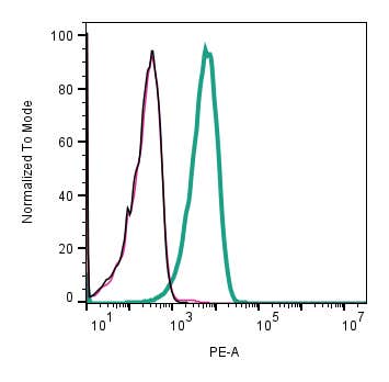 A Mouse Monoclonal Antibody to KCNN4 (KCa3.1, SK4) Channel
- Product Code: ALM-051-PE
- Short Description: A Mouse Monoclonal Antibody to KCNN4 (KCa3.1, SK4) Channel
- Host: Mouse
- Label: R-PE
- Reactivity: Human, Rat, Mouse
- Applications: Indirect flow cytometry, Live cell imaging, Western blot, Immunocytochemistry, Immunofluorescence, Immunohistochemistry
- Target: IKCa1, IK1, Intermediate conductance Ca2+-activated K+ channel protein 4, Gardos channel
-
 A Mouse Monoclonal Antibody to Rat Neurotrophin Receptor p75NTR
- Product Code: AN-170
- Short Description: A Mouse Monoclonal Antibody to Rat Neurotrophin Receptor p75NTR
- Host: Mouse
- Label: Unconjugated
- Reactivity: Rat
- Applications: Indirect flow cytometry, Western blot, Immunofluorescence, Immunohistochemistry
- Target: p75NTR, NGFR p75, TNFR superfamily member 16, TNFRSF16, CD271
-
 A Mouse Monoclonal Antibody to Rat Neurotrophin Receptor p75NTR Conjugated to the Fluorescent Dye FITC
- Product Code: AN-170-F
- Short Description: A Mouse Monoclonal Antibody to Rat Neurotrophin Receptor p75NTR Conjugated to the Fluorescent Dye FITC
- Host: Mouse
- Label: FITC
- Reactivity: Rat
- Applications: Flow cytometry
- Target: p75NTR, NGFR p75, TNFR superfamily member 16, TNFRSF16, CD271
-

 A biotinylated mouse NGF 2.5S for imaging and sensitive detection of TrkA and p75NTR receptors
- Product Code: N-240-B
- Short Description: A biotinylated mouse NGF 2.5S for imaging and sensitive detection of TrkA and p75NTR receptors
- Label: Biotin
- Applications: Cell Culture, Survival assay, Fluorescence staining, Live cell imaging, Immunofluorescence, Neurite outgrowth assay
- MW: ~26 kDa
-
 A Mouse Monoclonal Antibody to NGF and proNGF Designed to Neutralize their Biological Functions
- Product Code: ALM-006
- Short Description: A Mouse Monoclonal Antibody to NGF and proNGF Designed to Neutralize their Biological Functions
- Host: Mouse
- Label: Unconjugated
- Reactivity: Human, Mouse
- Applications: Indirect ELISA, Neutralization
- Target: Nerve growth factor
-
 A Blocking Peptide for Anti-MRS2 Antibody
- Product Code: BLP-NT148
- Short Description: A Blocking Peptide for Anti-MRS2 Antibody
- Applications: Western blot, Immunohistochemistry
- Target: MRS2
-
 A potent antagonist selective for the M4 muscarinic acetylcholine receptor (M4 mAChR) and a non-selective antagonist for several α1- and α2-adrenoceptor subtypes
- Product Code: M-140
- Short Description: A potent antagonist selective for the M4 muscarinic acetylcholine receptor (M4 mAChR) and a non-selective antagonist for several α1- and α2-adrenoceptor subtypes
- Applications: Calcium imaging assay
- MW: 7379 Da
-
 A negative allosteric modulator (NAM) of the M1 Muscarinic Receptor
- Product Code: STM-200
- Short Description: A negative allosteric modulator (NAM) of the M1 Muscarinic Receptor
- Applications: Electrophysiology
- CAS No.: 135541-77-4
- MW: 7472.5 Da
-
 A Blocking Peptide for Anti-N-Cadherin Antibody
- Product Code: BLP-NR082
- Short Description: A Blocking Peptide for Anti-N-Cadherin Antibody
- Applications: Western blot, Immunohistochemistry
- Target: CDH2
Loading...