-

 A Rabbit Polyclonal Antibody to KCNK16 Channel
- Product Code: APC-170
- Short Description: A Rabbit Polyclonal Antibody to KCNK16 Channel
- Host: Rabbit
- Label: Unconjugated
- Reactivity: Human, Rat, Mouse
- Applications: Western blot, Immunohistochemistry
- Target: Potassium two pore domain channel subfamily K member 16, TWIK-related alkaline pH-activated K+ channel 1, K2P16.1
-
 A Rabbit Polyclonal Antibody to KCNK18 Channel
- Product Code: APC-122
- Short Description: A Rabbit Polyclonal Antibody to KCNK18 Channel
- Host: Rabbit
- Label: Unconjugated
- Reactivity: Rat, Mouse
- Applications: Live cell imaging, Western blot, Immunocytochemistry, Immunofluorescence, Immunohistochemistry
- Target: Potassium channel subfamily K member 18, TWIK-related spinal cord K+ channel, K2P18.1
-
 A Rabbit Polyclonal Antibody to KCNK9 Channel
- Product Code: APC-044
- Short Description: A Rabbit Polyclonal Antibody to KCNK9 Channel
- Host: Rabbit
- Label: Unconjugated
- Reactivity: Human, Mouse
- Applications: Western blot, Immunofluorescence, Immunohistochemistry
- Target: Potassium channel subfamily K member 9, K2P9.1, TWIK-related acid-sensitive K+ channel 3, Two pore K+ channel KT3.2
-
 A Rabbit Polyclonal Antibody to KCNMA1 (KCa1.1) Channel
- Product Code: APC-151
- Short Description: A Rabbit Polyclonal Antibody to KCNMA1 (KCa1.1) Channel
- Host: Rabbit
- Label: Unconjugated
- Reactivity: Human, Rat, Mouse
- Applications: Indirect flow cytometry, Live cell imaging, Western blot, Immunofluorescence, Immunohistochemistry
- Target: Large conductance calcium-activated potassium channel subfamily M subunit alpha-1, BKCa alpha, Maxi K+, Slo1
-
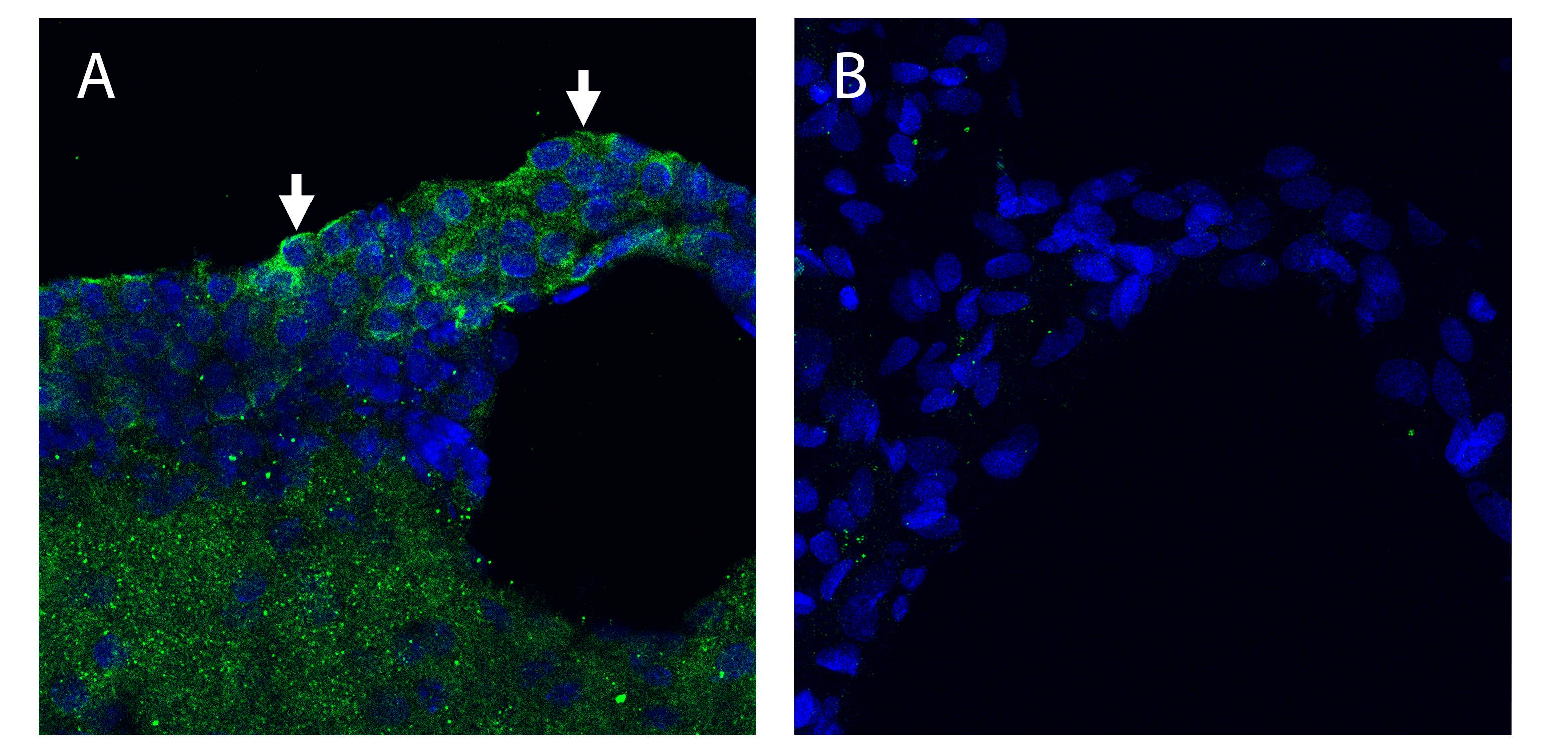 A Rabbit Polyclonal Antibody to KCNQ1 (KV7.1) Channel
- Product Code: APC-168
- Short Description: A Rabbit Polyclonal Antibody to KCNQ1 (KV7.1) Channel
- Host: Rabbit
- Label: Unconjugated
- Reactivity: Human, Rat, Mouse
- Applications: Indirect flow cytometry, Live cell imaging, Western blot, Immunocytochemistry, Immunofluorescence
- Target: KV7.1, Voltage-gated potassium channel QKT subfamily member 1, KvLQT1, KCNA9
-
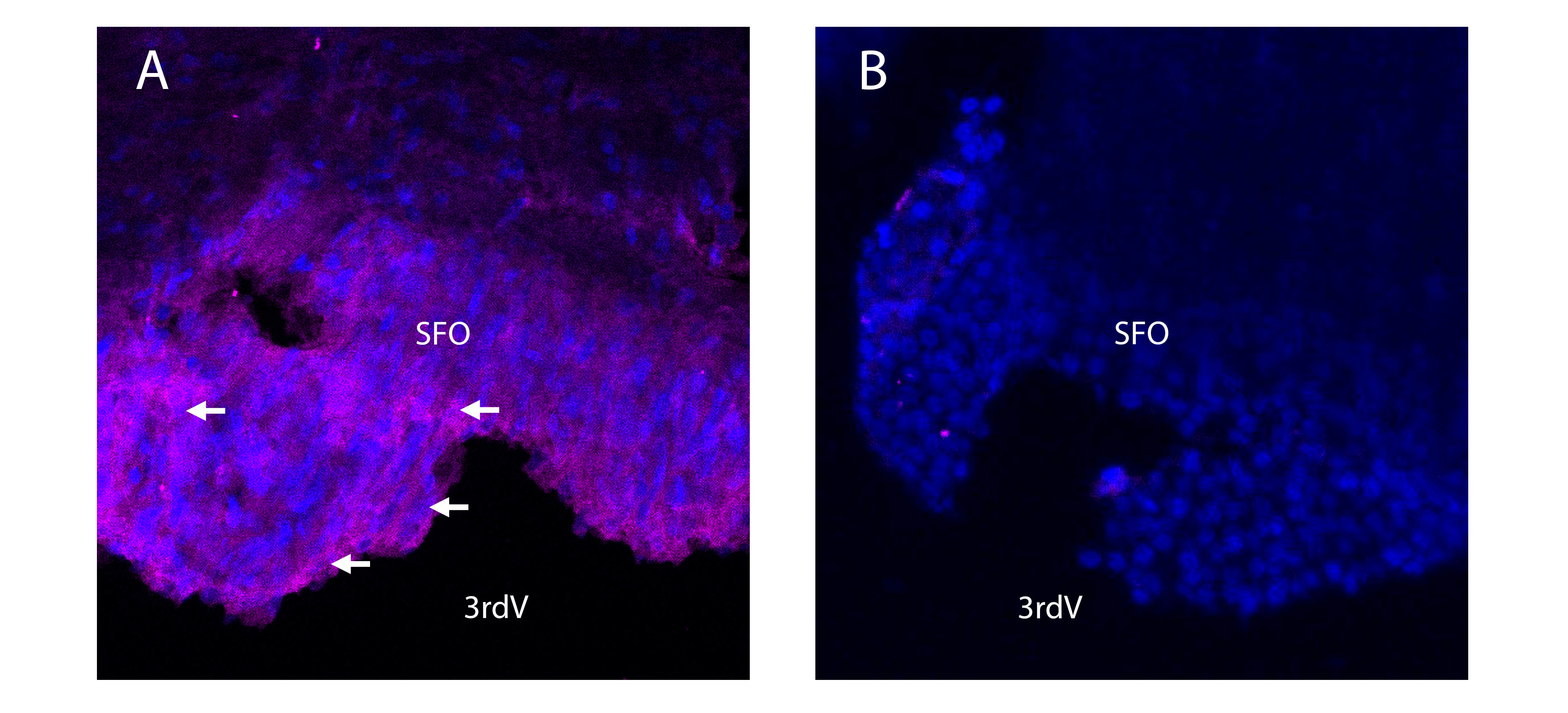 A Rabbit Polyclonal Antibody to KV7.1 (extracellular) conjugated to the fluorescent dye ATTO Fluor-647N.
- Product Code: APC-168-FRN
- Short Description: A Rabbit Polyclonal Antibody to KV7.1 (extracellular) conjugated to the fluorescent dye ATTO Fluor-647N.
- Host: Rabbit
- Label: ATTO 647N
- Reactivity: Human, Rat, Mouse
- Applications: Live cell imaging, Flow cytometry, Immunohistochemistry
- Target: Kcnq1
-
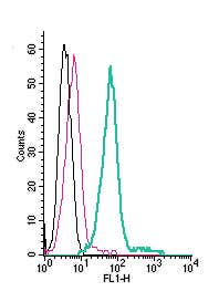 A Rabbit Polyclonal Antibody to KV7.1 (extracellular) conjugated to the fluorescent dye FITC
- Product Code: APC-168-F
- Short Description: A Rabbit Polyclonal Antibody to KV7.1 (extracellular) conjugated to the fluorescent dye FITC
- Host: Rabbit
- Label: FITC
- Reactivity: Human, Rat, Mouse
- Applications: Live cell imaging, Flow cytometry
- Target: KV7.1, Voltage-gated potassium channel QKT subfamily member 1, KvLQT1, KCNA9
-
 A Rabbit Polyclonal Antibody to KCNS1 (KV9.1) Channel
- Product Code: APC-131
- Short Description: A Rabbit Polyclonal Antibody to KCNS1 (KV9.1) Channel
- Host: Rabbit
- Label: Unconjugated
- Reactivity: Human, Rat, Mouse
- Applications: Western blot
- Target: Potassium voltage-gated channel subfamily S member 1
-
 A Rabbit Polyclonal Antibody to KCNS3 (KV9.3) Channel
- Product Code: APC-133
- Short Description: A Rabbit Polyclonal Antibody to KCNS3 (KV9.3) Channel
- Host: Rabbit
- Label: Unconjugated
- Reactivity: Human, Rat, Mouse
- Applications: Western blot, Immunofluorescence, Immunohistochemistry
- Target: Potassium voltage-gated channel subfamily S member 3
-
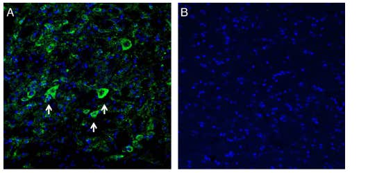 A Rabbit Polyclonal Antibody to Kir2.1 (KCNJ2) Channel
- Product Code: APC-159
- Short Description: A Rabbit Polyclonal Antibody to Kir2.1 (KCNJ2) Channel
- Host: Rabbit
- Label: Unconjugated
- Reactivity: Human, Rat, Mouse
- Applications: Indirect flow cytometry, Live cell imaging, Western blot, Immunocytochemistry, Immunofluorescence, Immunohistochemistry
- Target: Inward rectifier potassium channel 2, IRK1, HIRK1, LQT7, SQT3
Loading...