-

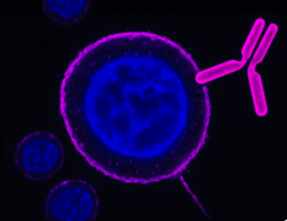
 A Rabbit Polyclonal Antibody to Piezo1 Channel
- Product Code: APC-087
- Short Description: A Rabbit Polyclonal Antibody to Piezo1 Channel
- Host: Rabbit
- Label: Unconjugated
- Reactivity: Rat, Mouse
- Applications: Indirect flow cytometry, Live cell imaging, Western blot, Immunofluorescence, Immunohistochemistry
- Target: Piezo-type mechanosensitive ion channel component 1, Protein FAM38A, Mib, Membrane protein induced by beta-amyloid treatment
-
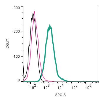 A Rabbit Polyclonal Antibody to Piezo1 (extracellular) conjugated to the fluorescent dye Allophycocyanin (APC)
- Product Code: APC-087-APC
- Short Description: A Rabbit Polyclonal Antibody to Piezo1 (extracellular) conjugated to the fluorescent dye Allophycocyanin (APC)
- Host: Rabbit
- Label: APC
- Reactivity: Rat, Mouse
- Applications: Live cell imaging, Flow cytometry
- Target: Piezo-type mechanosensitive ion channel component 1, Protein FAM38A, Mib, Membrane protein induced by beta-amyloid treatment
-
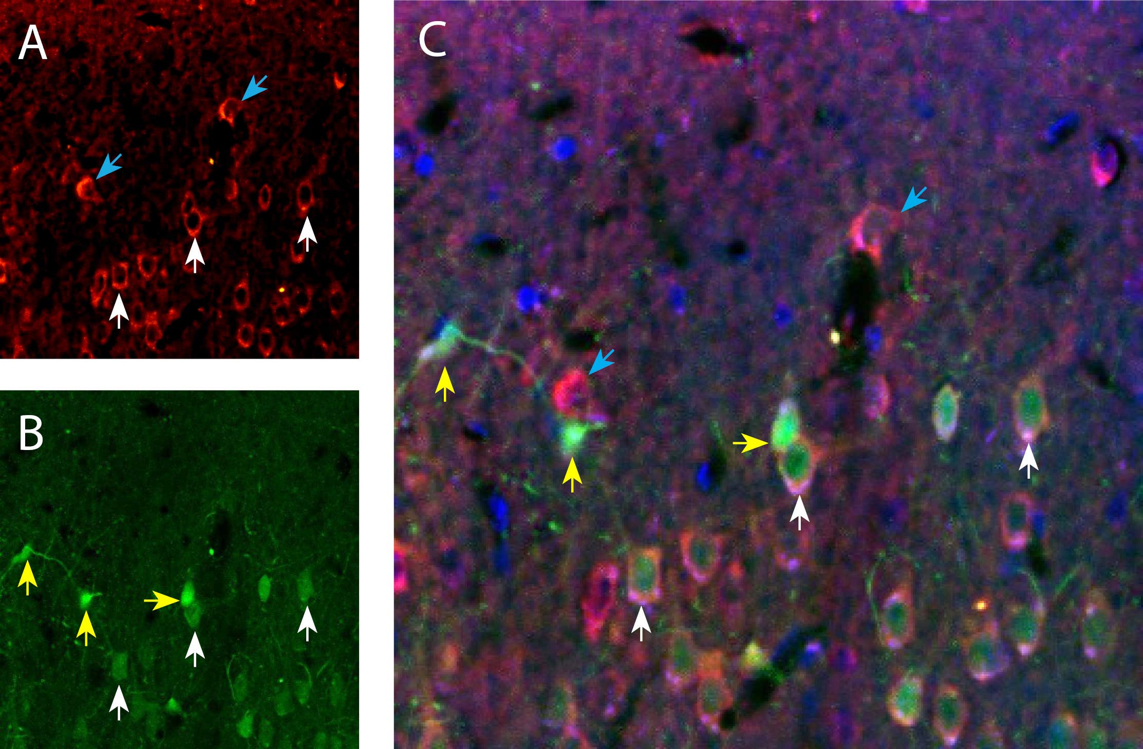 A Rabbit Polyclonal Antibody to Piezo1 (extracellular) conjugated to the fluorescent dye ATTO Fluor-550.
- Product Code: APC-087-AO
- Short Description: A Rabbit Polyclonal Antibody to Piezo1 (extracellular) conjugated to the fluorescent dye ATTO Fluor-550.
- Host: Rabbit
- Label: ATTO 550
- Reactivity: Rat, Mouse
- Applications: Live cell imaging, Flow cytometry, Immunohistochemistry
- Target: Piezo1
-
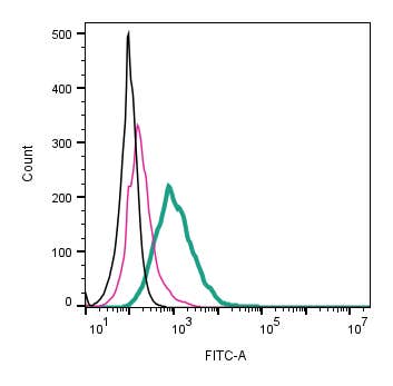 A Rabbit Polyclonal Antibody to Piezo1 (extracellular) conjugated to the fluorescent dye FITC
- Product Code: APC-087-F
- Short Description: A Rabbit Polyclonal Antibody to Piezo1 (extracellular) conjugated to the fluorescent dye FITC
- Host: Rabbit
- Label: FITC
- Reactivity: Rat, Mouse
- Applications: Live cell imaging, Flow cytometry
- Target: Piezo-type mechanosensitive ion channel component 1, Protein FAM38A, Mib, Membrane protein induced by beta-amyloid treatment
-
 A Rabbit Polyclonal antibody to Platelet-activating factor receptor (extracellular)
- Product Code: APR-055
- Short Description: A Rabbit Polyclonal antibody to Platelet-activating factor receptor (extracellular)
- Host: Rabbit
- Label: Unconjugated
- Reactivity: Human, Rat, Mouse
- Applications: Indirect flow cytometry, Live cell imaging, Western blot, Immunohistochemistry
- Target: PTAFR, PAFR
-
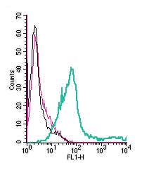 A Rabbit Polyclonal Antibody to Platelet-activating factor receptor (extracellular) conjugated to the fluorescent dye FITC
- Product Code: APR-055-F
- Short Description: A Rabbit Polyclonal Antibody to Platelet-activating factor receptor (extracellular) conjugated to the fluorescent dye FITC
- Host: Rabbit
- Label: FITC
- Reactivity: Human, Rat, Mouse
- Applications: Live cell imaging, Flow cytometry
- Target: PTAFR, PAFR
-
 A Rabbit Polyclonal Antibody to Plexin-A1
- Product Code: APR-081
- Short Description: A Rabbit Polyclonal Antibody to Plexin-A1
- Host: Rabbit
- Label: Unconjugated
- Reactivity: Human, Rat, Mouse
- Applications: Indirect flow cytometry, Live cell imaging, Western blot, Immunofluorescence, Immunohistochemistry
- Target: PLXNA1, PLXN1, Semaphorin receptor NOV
-
 A Rabbit Polyclonal Antibody to Plexin-A1 Conjugated to the Fluorescent Dye FITC
- Product Code: APR-081-F
- Short Description: A Rabbit Polyclonal Antibody to Plexin-A1 Conjugated to the Fluorescent Dye FITC
- Host: Rabbit
- Label: FITC
- Reactivity: Human, Rat, Mouse
- Applications: Live cell imaging, Flow cytometry
- Target: PLXNA1, PLXN1, Semaphorin receptor NOV
-
 A Rabbit Polyclonal Antibody to Plexin-A2
- Product Code: APR-082
- Short Description: A Rabbit Polyclonal Antibody to Plexin-A2
- Host: Rabbit
- Label: Unconjugated
- Reactivity: Human, Rat, Mouse
- Applications: Indirect flow cytometry, Live cell imaging, Western blot, Immunofluorescence, Immunohistochemistry
- Target: PLXNA2, PLXN2, Semaphorin receptor OCT
-
 A Rabbit Polyclonal Antibody to Plexin-A3
- Product Code: APR-093
- Short Description: A Rabbit Polyclonal Antibody to Plexin-A3
- Host: Rabbit
- Label: Unconjugated
- Reactivity: Human, Rat, Mouse
- Applications: Indirect flow cytometry, Live cell imaging, Western blot, Immunofluorescence, Immunohistochemistry
- Target: PLXNA3, PLXN3, Semaphorin receptor SEX
Loading...