-
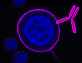
 A Mouse Monoclonal Antibody to GIRK1 (Kir3.1) Channel
- Product Code: ALM-031
- Short Description: A Mouse Monoclonal Antibody to GIRK1 (Kir3.1) Channel
- Host: Mouse
- Label: Unconjugated
- Reactivity: Human, Rat
- Applications: Live cell imaging, Western blot, Immunocytochemistry, Immunofluorescence
- Target: KCNJ3, G protein-activated inward rectifier potassium channel 1
-
 A Mouse Monoclonal Antibody to Human Orai1 Channel
- Product Code: ALM-025
- Short Description: A Mouse Monoclonal Antibody to Human Orai1 Channel
- Host: Mouse
- Label: Unconjugated
- Reactivity: Human
- Applications: Indirect flow cytometry, Live cell imaging, Western blot, Immunocytochemistry, Immunofluorescence
- Target: TMEM142A, CRACM1, Calcium release-activated calcium channel protein 1
-
 A Mouse Monoclonal Antibody to Human Orai1 Channel Conjugated to the Fluorescent Dye ATTO-633
- Product Code: ALM-025-FR
- Short Description: A Mouse Monoclonal Antibody to Human Orai1 Channel Conjugated to the Fluorescent Dye ATTO-633
- Host: Mouse
- Label: ATTO 633
- Reactivity: Human
- Applications: Live cell imaging, Flow cytometry
- Target: TMEM142A, CRACM1, Calcium release-activated calcium channel protein 1
-
 A Mouse Monoclonal Antibody to Human Orai1 Channel Conjugated to Biotin
- Product Code: ALM-025-B
- Short Description: A Mouse Monoclonal Antibody to Human Orai1 Channel Conjugated to Biotin
- Host: Mouse
- Label: Biotin
- Reactivity: Human
- Applications: Western blot, Flow cytometry
- Target: TMEM142A, CRACM1, Calcium release-activated calcium channel protein 1
-
 A Mouse Monoclonal Antibody to KCNN4 (KCa3.1, SK4) Channel
- Product Code: ALM-051
- Short Description: A Mouse Monoclonal Antibody to KCNN4 (KCa3.1, SK4) Channel
- Host: Mouse
- Label: Unconjugated
- Reactivity: Human, Rat, Mouse
- Applications: Indirect flow cytometry, Live cell imaging, Western blot, Immunocytochemistry, Immunofluorescence, Immunoprecipitation, Immunohistochemistry
- Target: IKCa1, IK1, Intermediate conductance Ca2+-activated K+ channel protein 4, Gardos channel
-
 A Mouse Monoclonal Antibody to KCNN4 (KCa3.1, SK4) Channel Conjugated to the Fluorescent Dye ATTO-488
- Product Code: ALM-051-AG
- Short Description: A Mouse Monoclonal Antibody to KCNN4 (KCa3.1, SK4) Channel Conjugated to the Fluorescent Dye ATTO-488
- Host: Mouse
- Label: ATTO 488
- Reactivity: Human, Rat, Mouse
- Applications: Live cell imaging, Immunocytochemistry, Immunofluorescence, Flow cytometry
- Target: IKCa1, IK1, Intermediate conductance Ca2+-activated K+ channel protein 4, Gardos channel
-
 A Mouse Monoclonal Antibody to KCNN4 (KCa3.1, SK4) Channel Conjugated to Biotin
- Product Code: ALM-051-B
- Short Description: A Mouse Monoclonal Antibody to KCNN4 (KCa3.1, SK4) Channel Conjugated to Biotin
- Host: Mouse
- Label: Biotin
- Reactivity: Human, Rat, Mouse
- Applications: Live cell imaging, Immunocytochemistry, Immunofluorescence, Flow cytometry
- Target: IKCa1, IK1, Intermediate conductance Ca2+-activated K+ channel protein 4, Gardos channel
-
 A Mouse Monoclonal Antibody to KCa3.1 Channel Conjugated to the Fluorescent Dye FITC
- Product Code: ALM-051-F
- Short Description: A Mouse Monoclonal Antibody to KCa3.1 Channel Conjugated to the Fluorescent Dye FITC
- Host: Mouse
- Label: FITC
- Reactivity: Human, Rat, Mouse
- Applications: Live cell imaging, Flow cytometry
- Target: IKCa1, IK1, Intermediate conductance Ca2+-activated K+ channel protein 4, Gardos channel
-
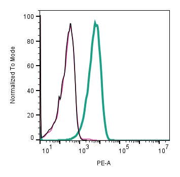 A Mouse Monoclonal Antibody to KCNN4 (KCa3.1, SK4) Channel
- Product Code: ALM-051-PE
- Short Description: A Mouse Monoclonal Antibody to KCNN4 (KCa3.1, SK4) Channel
- Host: Mouse
- Label: R-PE
- Reactivity: Human, Rat, Mouse
- Applications: Indirect flow cytometry, Live cell imaging, Western blot, Immunocytochemistry, Immunofluorescence, Immunohistochemistry
- Target: IKCa1, IK1, Intermediate conductance Ca2+-activated K+ channel protein 4, Gardos channel
-
 A Mouse Monoclonal Antibody to Rat Neurotrophin Receptor p75NTR
- Product Code: AN-170
- Short Description: A Mouse Monoclonal Antibody to Rat Neurotrophin Receptor p75NTR
- Host: Mouse
- Label: Unconjugated
- Reactivity: Rat
- Applications: Indirect flow cytometry, Western blot, Immunofluorescence, Immunohistochemistry
- Target: p75NTR, NGFR p75, TNFR superfamily member 16, TNFRSF16, CD271
Loading...