-
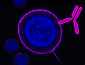
 A Rabbit Polyclonal Antibody to KV1.3 (KCNA3) Channel Conjugated to Biotin
- Product Code: APC-101-B
- Short Description: A Rabbit Polyclonal Antibody to KV1.3 (KCNA3) Channel Conjugated to Biotin
- Host: Rabbit
- Label: Biotin
- Reactivity: Human, Rat, Mouse
- Applications: Western blot, Immunofluorescence, Flow cytometry, Immunohistochemistry
- Target: Potassium voltage-gated channel subfamily A member 3
-
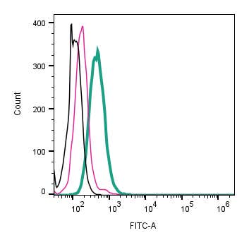 A Rabbit Polyclonal Antibody to KV1.3 (KCNA3) Channel Conjugated to the Fluorescent Dye FITC
- Product Code: APC-101-F
- Short Description: A Rabbit Polyclonal Antibody to KV1.3 (KCNA3) Channel Conjugated to the Fluorescent Dye FITC
- Host: Rabbit
- Label: FITC
- Reactivity: Human, Rat, Mouse
- Applications: Live cell imaging, Immunocytochemistry, Immunofluorescence, Flow cytometry
- Target: Potassium voltage-gated channel subfamily A member 3
-
 A Rabbit Polyclonal Antibody to KV1.3 (KCNA3) Channel Conjugated to the Fluorescent Dye Phycoerythrin
- Product Code: APC-101-PE
- Short Description: A Rabbit Polyclonal Antibody to KV1.3 (KCNA3) Channel Conjugated to the Fluorescent Dye Phycoerythrin
- Host: Rabbit
- Label: R-PE
- Reactivity: Human, Rat, Mouse
- Applications: Live cell imaging, Flow cytometry
- Target: Potassium voltage-gated channel subfamily A member 3
-
 A Rabbit Polyclonal Antibody to KV1.4 Channel
- Product Code: APC-167
- Short Description: A Rabbit Polyclonal Antibody to KV1.4 Channel
- Host: Rabbit
- Label: Unconjugated
- Reactivity: Rat, Mouse
- Applications: Live cell imaging, Western blot, Immunocytochemistry, Immunofluorescence, Immunohistochemistry
- Target: Potassium voltage-gated channel subfamily A member 4, KCNA4
-
 A Rabbit Polyclonal Antibody to KV1.5 (KCNA5) Channel
- Product Code: APC-150
- Short Description: A Rabbit Polyclonal Antibody to KV1.5 (KCNA5) Channel
- Host: Rabbit
- Label: Unconjugated
- Reactivity: Human, Mouse
- Applications: Indirect flow cytometry, Live cell imaging, Western blot, Immunocytochemistry, Immunofluorescence, Immunohistochemistry
- Target: Potassium voltage-gated channel subfamily A member 5
-
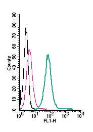 A Rabbit Polyclonal Antibody to KV1.5 (extracellular) conjugated to the fluorescent dye FITC
- Product Code: APC-150-F
- Short Description: A Rabbit Polyclonal Antibody to KV1.5 (extracellular) conjugated to the fluorescent dye FITC
- Host: Rabbit
- Label: FITC
- Reactivity: Human, Rat, Mouse
- Applications: Live cell imaging, Flow cytometry
- Target: Potassium voltage-gated channel subfamily A member 5
-
 A Rabbit Polyclonal antibody to L1CAM (extracellular)
- Product Code: ANR-047
- Short Description: A Rabbit Polyclonal antibody to L1CAM (extracellular)
- Host: Rabbit
- Label: Unconjugated
- Reactivity: Human, Rat, Mouse
- Applications: Indirect flow cytometry, Live cell imaging, Western blot, Immunohistochemistry
- Target: L1 Cell Adhesion Molecule, Neural Cell Adhesion Molecule L1, NCAM-L1, CAML1, CD171
-
 A Rabbit Polyclonal antibody to LAT2 (SLC7A8) (extracellular)
- Product Code: ANT-108
- Short Description: A Rabbit Polyclonal antibody to LAT2 (SLC7A8) (extracellular)
- Host: Rabbit
- Label: Unconjugated
- Reactivity: Human, Rat, Mouse
- Applications: Indirect flow cytometry, Live cell imaging, Western blot, Immunohistochemistry
- Target: Large Neutral Amino Acids Transporter Small Subunit 2, L-Type Amino Acid Transporter 2, Solute carrier family 7 member 8
-
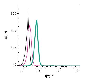 A Rabbit Polyclonal antibody to LAT2 (SLC7A8) (extracellular) conjugated to the fluorescent dye FITC
- Product Code: ANT-108-F
- Short Description: A Rabbit Polyclonal antibody to LAT2 (SLC7A8) (extracellular) conjugated to the fluorescent dye FITC
- Host: Rabbit
- Label: FITC
- Reactivity: Human, Rat, Mouse
- Applications: Live cell imaging, Flow cytometry
- Target: Large Neutral Amino Acids Transporter Small Subunit 2, L-Type Amino Acid Transporter 2, Solute carrier family 7 member 8
-
 A Rabbit Polyclonal Antibody to LAT3 Transporter
- Product Code: ANT-193
- Short Description: A Rabbit Polyclonal Antibody to LAT3 Transporter
- Host: Rabbit
- Label: Unconjugated
- Reactivity: Human, Rat, Mouse
- Applications: Western blot
- Target: Large neutral amino acids transporter small subunit 3, L-type amino acid transporter 3, Solute carrier family 43 member 1
Loading...