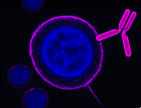
-
 A Rabbit Polyclonal antibody to C3aR1 (extracellular)
- Product Code: AAR-036
- Short Description: A Rabbit Polyclonal antibody to C3aR1 (extracellular)
- Host: Rabbit
- Label: Unconjugated
- Reactivity: Rat, Mouse
- Applications: Indirect flow cytometry, Live cell imaging, Western blot, Immunohistochemistry
- Target: Complement C3a Receptor 1, C3AR, C3a Anaphylatoxin Chemotactic Receptor
-
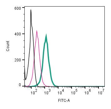 A Rabbit Polyclonal Antibody to C3aR1 (extracellular) conjugated to the fluorescent dye FITC
- Product Code: AAR-036-F
- Short Description: A Rabbit Polyclonal Antibody to C3aR1 (extracellular) conjugated to the fluorescent dye FITC
- Host: Rabbit
- Label: FITC
- Reactivity: Rat, Mouse
- Applications: Live cell imaging, Flow cytometry
- Target: Complement C3a Receptor 1, C3AR, C3a Anaphylatoxin Chemotactic Receptor
-
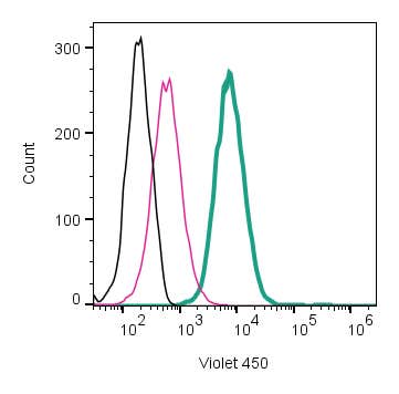 A Rabbit Polyclonal Antibody to C3aR1 (extracellular) conjugated to the fluorescent dye mFluor™ Violet 450 fluorophore.
- Product Code: AAR-036-V
- Short Description: A Rabbit Polyclonal Antibody to C3aR1 (extracellular) conjugated to the fluorescent dye mFluor™ Violet 450 fluorophore.
- Host: Rabbit
- Label: mFluor™ Violet 450
- Reactivity: Rat, Mouse
- Applications: Live cell imaging, Flow cytometry
- Target: Complement C3a Receptor 1, C3AR, C3a Anaphylatoxin Chemotactic Receptor
-
 A Rabbit Polyclonal antibody to C5aR2 (extracellular).
- Product Code: AAR-034
- Short Description: A Rabbit Polyclonal antibody to C5aR2 (extracellular).
- Host: Rabbit
- Label: Unconjugated
- Reactivity: Rat, Mouse
- Applications: Indirect flow cytometry, Western blot, Immunohistochemistry
- Target: Complement Component 5a Receptor 2, C5L2, C5a Anaphylatoxin Chemotactic Receptor 2, G Protein-Coupled Receptor 77, GPR77
-

 A Rabbit Polyclonal Antibody to CACNA2D1 (CaVα2δ1 Subunit)
- Product Code: ACC-015
- Short Description: A Rabbit Polyclonal Antibody to CACNA2D1 (CaVα2δ1 Subunit)
- Host: Rabbit
- Label: Unconjugated
- Reactivity: Human, Mouse
- Applications: Live cell imaging, Western blot, Immunocytochemistry, Immunofluorescence
- Target: Voltage-dependent Ca2+ channel subunit alpha-2/delta-1, L-type calcium channel alpha 2 polypeptide, Dihydropyridine-sensitive L-type calcium channel alpha-2/delta subunit
-
 A Rabbit Polyclonal Antibody to CACNA2D2 (CaVα2δ2) Subunit
- Product Code: ACC-102
- Short Description: A Rabbit Polyclonal Antibody to CACNA2D2 (CaVα2δ2) Subunit
- Host: Rabbit
- Label: Unconjugated
- Reactivity: Human, Rat, Mouse
- Applications: Indirect flow cytometry, Live cell imaging, Western blot, Immunofluorescence, Immunohistochemistry
- Target: Voltage-dependent calcium channel subunit alpha-2/delta-2
-
 A Rabbit Polyclonal Antibody to CACNA2D3 (CaVα2δ3) Subunit
- Product Code: ACC-103
- Short Description: A Rabbit Polyclonal Antibody to CACNA2D3 (CaVα2δ3) Subunit
- Host: Rabbit
- Label: Unconjugated
- Reactivity: Human, Rat, Mouse
- Applications: Live cell imaging, Western blot, Immunocytochemistry, Immunofluorescence, Immunohistochemistry
- Target: Voltage-dependent calcium channel subunit alpha-2/delta-3
-
 A Rabbit Polyclonal Antibody to CACNA2D3 (CaVα2δ3) Subunit Conjugated to the Fluorescent Dye ATTO-594
- Product Code: ACC-103-AR
- Short Description: A Rabbit Polyclonal Antibody to CACNA2D3 (CaVα2δ3) Subunit Conjugated to the Fluorescent Dye ATTO-594
- Host: Rabbit
- Label: ATTO 594
- Reactivity: Human, Rat, Mouse
- Applications: Live cell imaging, Immunocytochemistry, Immunofluorescence, Immunohistochemistry
- Target: Voltage-dependent calcium channel subunit alpha-2/delta-3
-
 A Rabbit Polyclonal Antibody to CACNA2D4 (CaVα2δ4) Subunit
- Product Code: ACC-104
- Short Description: A Rabbit Polyclonal Antibody to CACNA2D4 (CaVα2δ4) Subunit
- Host: Rabbit
- Label: Unconjugated
- Reactivity: Rat, Mouse
- Applications: Live cell imaging, Western blot, Immunocytochemistry, Immunofluorescence, Immunohistochemistry
- Target: Voltage-dependent calcium channel subunit alpha-2/delta-4, RCD4
-
 A Rabbit Polyclonal Antibody to CACNA2D4 (CaVα2δ4) Subunit Conjugated to the Fluorescent Dye ATTO-594
- Product Code: ACC-104-AR
- Short Description: A Rabbit Polyclonal Antibody to CACNA2D4 (CaVα2δ4) Subunit Conjugated to the Fluorescent Dye ATTO-594
- Host: Rabbit
- Label: ATTO 594
- Reactivity: Rat, Mouse
- Applications: Live cell imaging, Immunocytochemistry, Immunofluorescence
- Target: Voltage-dependent calcium channel subunit alpha-2/delta-4, RCD4
Loading...