Neurotrophin-Based Protocols
Conjugated Neurotrophin-based Live-cell Flow Cytometry
Live-cell flow cytometry using fluorophore- or biotin-conjugated neurotrophins enables quantitative analysis of their receptors in living cells. These highly specific probes allow sensitive detection of cell surface receptors while preserving cell viability. This protocol describes live-cell staining with conjugated neurotrophins for accurate receptor detection and flow cytometric analysis.
Materials
- Conjugated neurotrophin of choice (fluorophore- or biotin-labeled).
- Dulbecco’s Phosphate Buffered Saline (PBS) (02-023-1A, Sartorius).
- Streptavidin-labeled fluorophore (S21374, Invitrogen).
- CAS-Block™ (008120, ThermoFisher Scientific).
Procedure
Step 1 – Reagent Preparation
1.1 Dilute the conjugated neurotrophin according to the instructions provided in its Certificate of Analysis.
1.2 For biotin-conjugated neurotrophins, prepare the streptavidin-conjugated fluorophore according to the manufacturer’s instructions.
- Protect all fluorescent reagents from light during preparation and throughout the staining procedure to minimize photobleaching.
Step 2 – CAS-Block
CAS-Block is a universal blocking reagent for immunochemistry methods. It reduces non-specific binding and background staining, eliminating the need for species-specific serum blockers.
2.1 Incubate cells with CAS-Block for 30 minutes at 37°C.
Step 3 – Preparation of Cells
3.1 Transfer 1 × 10⁶ cells into a microcentrifuge (Eppendorf) tube and centrifuge for 5 minutes at 200 x g.
3.2 Carefully discard the supernatant. Resuspend the cell pellet in 1 mL of PBS by gentle pipetting and centrifuge for 5 minutes at 200 x g.
3.3 Carefully discard the supernatant.
- Minimize shear stress during pipetting to preserve cell viability and maintain staining integrity.
Step 4 – Neurotrophin Incubation
This step allows the conjugated neurotrophin to bind selectively to its receptor on the cell surface.
4.1 Add the prepared neurotrophin solution to the cells and gently resuspend by pipetting.
4.2 Incubate the cells for 30–60 minutes at 37°C in a cell culture incubator.
- Optimize the neurotrophin concentration and incubation time according to the target receptor and cell type.
- Mix gently to avoid mechanical stress and maintain cell viability.
Step 5 – Streptavidin-conjugated Fluorophore Incubation (for Biotin-conjugated Neurotrophins Only)
A streptavidin-conjugated fluorophore is used to detect and amplify the signal from biotinylated neurotrophins.
5.1 Wash the cells three times with PBS: resuspend the cell pellet in 1 mL of PBS by gentle pipetting and centrifuge for 5 minutes at 200 × g. Carefully discard the supernatant after each wash.
5.2 Add the streptavidin-conjugated fluorophore to the cells and gently resuspend.
5.3 Incubate for 15-30 minutes at 37°C in a cell culture incubator.
Step 6 – Washing
This step removes unbound streptavidin and excess conjugated neurotrophin, reducing background fluorescence.
6.1 Centrifuge the cells for 5 minutes at 200 x g and carefully discard the supernatant.
6.2 Resuspend the cell pellet in 1 mL of PBS, then centrifuge for 5 minutes at 200 x g.
6.3 Carefully discard the supernatant and resuspend the cells in 1 mL of PBS.
6.4 Filter the cell suspension appropriately before flow cytometry analysis.
Troubleshooting
| Problem | Possible Cause | Solution |
|---|---|---|
| Low signal | Fluorophore degradation; low receptor expression; suboptimal neurotrophin concentration | Verify proper storage and reconstitution of the neurotrophin; confirm target receptor expression in the selected cell line; optimize the neurotrophin concentration |
| High background | Excess neurotrophin; insufficient washing | Reduce neurotrophin concentration and/or incubation time; increase the number or duration of washing steps |
| Photobleaching | Prolonged light exposure | Minimize light exposure; protect stained cells from light and keep them in the dark until analysis |
| Inaccurate signal interpretation | Lack of proper controls | Include appropriate negative controls such as unstained cells, cells treated with unlabeled neurotrophin, and a streptavidin-only control (for biotin-conjugated neurotrophins) |
Example Data
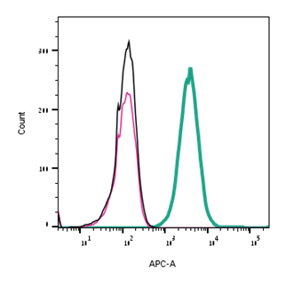
Direct flow cytometry of NGF expression in live intact rat PC-12 cells.
___ PC-12 cells.
___ PC-12 cells + 10 nM NGF (#N-240).
___ PC-12 cells + 10 nM NGF-ATTO Fluor-647N (#N-240-FRN).