Neurotrophin-Based Protocols
Immunofluorescence of Frozen Sections Using Conjugated Neurotrophins
Neurotrophins conjugated to fluorophores or biotin are valuable tools for the specific detection of target receptors in tissue sections. These labeled ligands enable the visualization and co-localization of cell membrane receptors in frozen tissue preparations. The following protocol describes the use of conjugated neurotrophins for tissue labeling, with an emphasis on precision, reproducibility, and optimal imaging outcomes.
Materials
- Conjugated neurotrophin of choice (fluorophore- or biotin-labeled).
- Dulbecco’s Phosphate Buffered Saline (PBS) (02-023-1A, Sartorius).
- Streptavidin-labeled fluorophore (S21374, Invitrogen).
- CAS-Block™ (008120, ThermoFisher Scientific).
- Hoechst 33342 (H3570, ThermoFisher Scientific).
- Mouse C57 Dorsal Root Ganglion Frozen Sections (MF-240-C57, Zyagen).
- Epredia™ Immu-Mount™ (9990402, ThermoFisher Scientific).
Procedure
Step 1 – Preparation of Tissue Sections
When preparing sections, use gentle to mild fixation methods to preserve the receptor structure and ensure proper neurotrophin binding.
1.1 Remove the slide box from the freezer without opening it and allow it to equilibrate to room temperature (RT) for at least 30 minutes. This step is essential to prevent condensation from forming on the slides.
1.2 Open the slide box and allow the slides to equilibrate at RT for an additional 10-15 minutes.
1.3 Fix the tissue sections with 4% paraformaldehyde (PFA) for 10 minutes at RT.
1.4 Wash the sections once with PBS (1X) to remove debris and rehydrate the tissue.
Step 2 – CAS-Block
CAS-Block is a universal blocking reagent for immunochemistry methods. It reduces non-specific binding and background staining, eliminating the need for species-specific serum blockers.
2.1 Apply a sufficient amount of CAS-Block to cover the frozen section.
- Incubate 30 minutes at RT.
Step 3 – Neurotrophin Incubation
This step allows the conjugated neurotrophin to selectively bind to its receptor within the tissue.
3.1 Dilute the conjugated neurotrophin according to the instructions provided in the Certificate of Analysis.
3.2 Apply the diluted neurotrophin to the tissue sections and place the slides in a humidified chamber.
- Incubate the sections for 1 hour at 37°C or overnight at 4°C.
- Optimize the neurotrophin concentration and incubation time for each tissue type.
- Protect fluorescent reagents from light to minimize photobleaching.
- Include appropriate controls: a control lacking the conjugated neurotrophin and an unlabeled neurotrophin control.
Step 4 – Streptavidin-conjugated Fluorophore Incubation (for Biotin-conjugated Neurotrophins Only)
A streptavidin-conjugated fluorophore is used to detect and amplify the signal from biotinylated neurotrophins.
4.1 Wash the tissue sections with PBS (1X) three times, for 5 min each, to remove unbound neurotrophin.
4.2 Apply the streptavidin-conjugated fluorophore to the tissue sections.
- Incubate 30 minutes at RT.
Step 5 – Nuclear Counterstaining (Optional)
This step provides cellular context by labeling cell nuclei.
5.1 Wash the tissue sections with PBS (1X) three times, for 5 min each.
5.2 Prepare Hoechst 33342 (2 µg/mL in PBS) and apply to the tissue sections.
- Incubate 10 minutes at RT.
5.3 Rinse the sections three times with PBS (1X).
Step 6 – Mounting and Imaging
6.1 Apply ~3 drops of mounting medium (e.g., Immu-Mount) onto each slide and affix a coverslip.
- Allow slides to dry for ≥30 minutes at RT.
6.2 Examine samples by fluorescence microscopy.
Troubleshooting
| Problem | Possible Cause | Solution |
|---|---|---|
| Low signal | Fluorophore degradation; low receptor expression; suboptimal neurotrophin concentration; insufficient incubation time | Verify proper storage and reconstitution of the neurotrophin; confirm receptor expression in the selected tissue; optimize neurotrophin concentration and incubation time |
| High background | Excess neurotrophin; insufficient washing | Reduce neurotrophin concentration and/or incubation time; increase the number or duration of washing steps |
| Photobleaching | Prolonged light exposure | Minimize light exposure; protect stained cells from light and keep them in the dark until analysis |
| Non-specific staining | Excessive fixation; unintended permeabilization; insufficient blocking | Reduce fixation strength or duration; avoid permeabilization when detecting surface receptors; extend the blocking duration |
Example Data
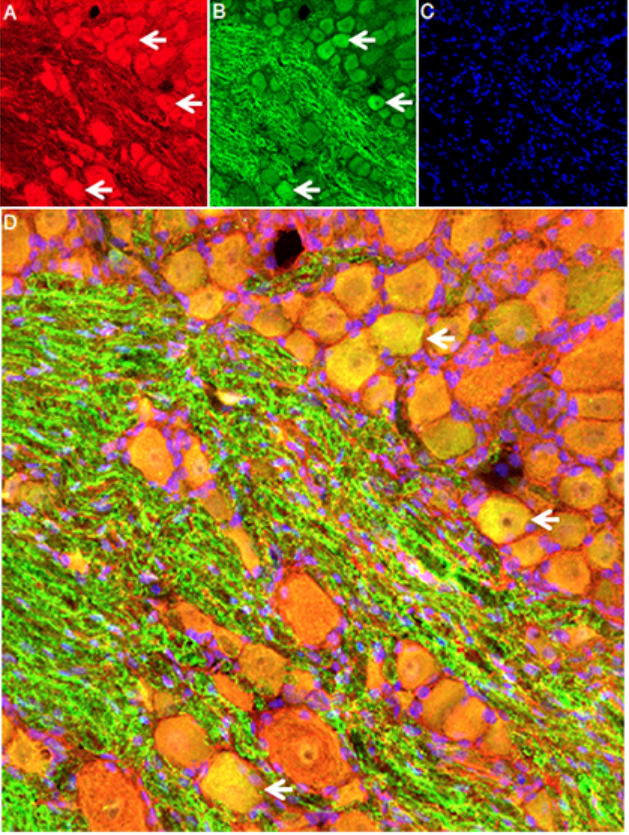
NGF co-localized with the p75 NGF Receptor (p75NTR) in rat DRG frozen tissue.
A) Fixed rat DRG sections were incubated with 0.1 µM of NGF-ATTO Fluor-647N (N-240-FRN) for 1h at 37°C. B) The same sections were stained with Anti-p75 NGF Receptor (extracellular)-ATTO Fluor-488 Antibody (#ANT-007-AG). C) The cell nuclei were stained with Hoechst 33342 (blue). D) Merging of the images A-C revealed co-staining of NGF-ATTO Fluor-647N, and p75NTR (arrows) in DRG cells.