Neurotrophin-Based Protocols
Live-Cell Imaging Using Conjugated Neurotrophins
Conjugated neurotrophins serve as selective probes for live-cell imaging and biodetection. These labeled ligands enable the visualization of receptors in living cells, facilitating the analysis of receptor localization, dynamics, and binding kinetics. The following protocol describes an optimized procedure for the use of conjugated neurotrophins in live-cell assays, designed to ensure reproducible results and reliable imaging performance.
Materials
- Conjugated neurotrophin of choice (fluorophore- or biotin-labeled).
- Dulbecco’s Phosphate Buffered Saline (PBS) (02-023-1A, Sartorius).
- Streptavidin-labeled fluorophore (S21374, Invitrogen).
- CAS-Block™ (008120, ThermoFisher Scientific).
Procedure
Step 1 – Reagent Preparation
1.1 Dilute the conjugated neurotrophin according to the instructions provided in its Certificate of Analysis.
1.2 For biotin-conjugated neurotrophins, prepare the streptavidin-conjugated fluorophore according to the manufacturer’s instructions.
- Protect all fluorescent reagents from light during preparation and throughout the staining procedure to minimize photobleaching.
Step 2 – CAS-Block
CAS-Block is a universal blocking reagent for immunochemistry methods. It reduces non-specific binding and background staining, eliminating the need for species-specific serum blockers.
2.1 Incubate the cells with CAS-Block for 30 minutes at 37°C.
Step 3 – Neurotrophin Incubation
This step allows the conjugated neurotrophin to bind selectively to its receptor on the cell surface.
3.1 Add the prepared neurotrophin solution to the adherent cells in imaging dishes.
3.2 Incubate the cells for 10-60 minutes at 37°C in a cell culture incubator.
- Optimize the neurotrophin concentration and incubation time according to the target receptor and cell type.
- Ensure the cells remain fully covered with the neurotrophin solution during incubation.
Step 4 – Streptavidin-conjugated Fluorophore Incubation (for Biotin-conjugated Neurotrophins Only)
A streptavidin-conjugated fluorophore is used to detect and amplify the signal from biotinylated neurotrophins.
4.1 Wash the cells three times with PBS for 5 minutes each.
4.2 Apply the streptavidin-conjugated fluorophore to the cells.
- Incubate 15-30 minutes at 37°C in an incubator.
Step 5 – Washing and Imaging
This step removes unbound conjugated streptavidin and excess conjugated neurotrophin, reducing background fluorescence.
5.1 Wash the cells three times with PBS: gently add PBS to the culture dish, swirl carefully, and remove the solution without disturbing the cells.
5.2 Image the cells using a microscope optimized for live-cell imaging.
Troubleshooting
| Problem | Possible Cause | Solution |
|---|---|---|
| Low signal | Fluorophore degradation; low target receptor expression; suboptimal neurotrophin concentration | Verify proper storage and reconstitution of the neurotrophin; confirm target receptor expression in the selected cell line; optimize neurotrophin concentration |
| High background | Excess neurotrophin; insufficient washing | Reduce neurotrophin concentration and/or incubation time; increase the number or duration of washing steps |
| Photobleaching | Prolonged light exposure | Minimize light exposure; protect stained cells from light and keep them in the dark until analysis |
| Inaccurate signal interpretation | Lack of appropriate controls | Include appropriate negative controls such as unstained cells, cells treated with unlabeled neurotrophin, and a streptavidin-only control (for biotin-conjugated neurotrophins) |
Example Data
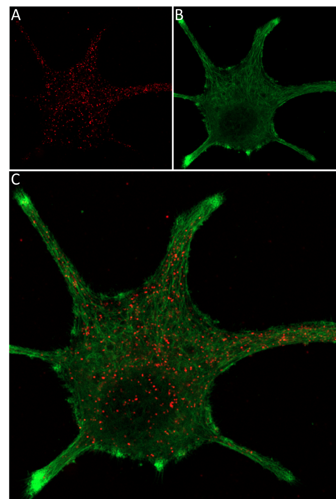
Live-cell imaging of mouse NGF 2.5S-ATTO Fluor-647N in differentiated PC-12 cells.
A) Neurite outgrowth was induced in PC-12 cells by 8 days of exposure to 100 ng/ml mouse NGF 2.5S-ATTO Fluor-647N (#N-240-FRN). B) The CellMask™ Actin 1X solution was applied for 30 minutes, resulting in green fluorescence, allowing visualization of the cellular membrane. C) Live-cell imaging of the differentiated PC-12 cell allowed observation of NGF distribution among the cell.