Toxin-Based Protocols
Multiplex Immunodetection Protocol
Suitable for Frozen Sections Using Conjugated Toxins and Unconjugated Antibodies Directed Against Extracellular Epitopes.
Venom-derived peptide toxins conjugated to fluorophores or biotin can be used for the specific detection of molecular targets in frozen tissue sections. These reagents enable visualization and co-localization of cell membrane proteins while preserving native tissue architecture. Protein localization is performed using immunofluorescence (IF) with antibodies directed against defined extracellular epitopes. In indirect IF, a fluorophore-conjugated secondary antibody binds to the primary antibody targeting the protein of interest. Although this approach requires additional steps compared with direct labeling of the primary antibody, it provides enhanced sensitivity through signal amplification, as multiple secondary antibodies can bind to a single primary antibody. This amplification may also increase background staining. The protocol below describes immunofluorescent labeling of frozen tissue sections using fluorophore- or biotin-conjugated peptide toxins in combination with primary antibodies directed against extracellular epitopes, with emphasis on reproducible staining and high-quality fluorescence imaging.
Materials
- Conjugated toxin of choice (fluorophore- or biotin-labeled).
- Primary antibody and secondary antibody validated for IF use.
- CAS-Block™ (008120, ThermoFisher Scientific).
- Dulbecco’s Phosphate Buffered Saline (PBS) (02-023-1A, Sartorius).
- Streptavidin-labeled fluorophore (for biotin-conjugated toxins) [S21374, Invitrogen].
- Hoechst 33342 (H3570, ThermoFisher Scientific).
- Epredia™ Immu-Mount™ (9990402, ThermoFisher Scientific).
Procedure
Step 1 – Preparation of Tissue Sections
1.1 Remove the slide box from the freezer (do not open) and allow it to equilibrate to room temperature (RT) for at least 30 minutes. This step is essential to prevent condensation from forming on the slides.
1.2 After opening the slide box, it is also recommended to leave slides at RT for an additional 10-15 minutes.
1.3 Fix the tissue sections with 4% paraformaldehyde for 10 minutes.
1.4 Wash the sections once with PBS (1X) to remove debris and rehydrate the tissue.
Step 2 – CAS-Block
CAS-Block is a universal blocking reagent for immunochemical methods. It reduces non-specific binding and background staining, eliminating the need for species-specific serum blockers.
2.1 Apply a sufficient amount of CAS-Block to cover the frozen section.
- Incubate 30 minutes at RT.
Step 3 – Primary Antibody Incubation
This step allows the unconjugated primary antibody to specifically identify its designated epitope within the tissue.
3.1 Dilute the unconjugated extracellular primary antibody according to the instructions provided in its Certificate of Analysis.
3.2 Apply the diluted antibody on the tissue sections and place in a humidified chamber.
- Incubate overnight at 4°C.
- Optimize antibody concentration for each tissue type.
- Include appropriate controls: negative and positive controls.
Step 4 – Secondary Antibody Incubation
This stage enables the conjugated secondary antibody to recognize the primary antibody, allowing for visualization of its target.
4.1 Wash the sections with PBS (1X) three times, for five minutes each, to eliminate any unbound primary antibody.
4.2 Apply the appropriate dilution of the secondary antibody.
- Incubate 30 minutes at RT.
- Protect fluorescent reagents from light to avoid photobleaching.
Step 5 – Toxin Incubation
This step enables the conjugated toxin to selectively bind to its molecular target within the tissue.
5.1 Wash the sections with PBS (1X) three times, for five minutes each, to eliminate any unbound secondary antibody.
5.2 Dilute the conjugated toxin according to the instructions provided in its Certificate of Analysis.
5.3 Apply the diluted toxin on the tissue sections and place in a humidified chamber.
- Incubate 1 hour at 37°C.
- Optimize toxin concentration and incubation time for each tissue type and target molecule.
- Protect fluorescent reagents from light to avoid photobleaching.
- Include appropriate controls: a control lacking the toxin and an unlabeled toxin control.
Step 6 – Streptavidin (for biotin-conjugated toxins only)
A streptavidin-fluorophore amplifies the signal from biotinylated toxins.
6.1 Wash sections with PBS (1X) three times, for five minutes each, to remove unbound toxin.
6.2 Apply the streptavidin-conjugated fluorophore.
- Incubate 30 minutes at RT.
Step 7 – Nuclear Counterstaining (Optional)
Provides cellular context by labeling nuclei.
7.1 Wash sections with PBS (1X) three times, for five minutes each.
7.2 For nuclear counterstaining: Apply Hoechst 33342 (2 μg/mL).
- Incubate 10 minutes at RT.
7.3 Wash sections briefly again, three times with PBS (1X).
Step 8 – Mounting and Imaging
8.1 Apply ~3 drops of mounting medium (e.g., Immu-Mount) onto each slide and affix a coverslip.
- Allow slides to dry for ≥30 minutes at RT.
Troubleshooting
| Problem | Possible Cause | Solution |
|---|---|---|
| Low signal | Fluorophore degradation; poor target expression; inappropriate toxin or antibody concentration | Verify storage and reconstitution of toxin/antibody; confirm target protein expression in the chosen cell line; optimize toxin/antibody concentration |
| High background | Excess toxin/antibody; insufficient washing | Reduce toxin/antibody concentration and/or incubation time; increase washing steps |
| Photobleaching | Prolonged light exposure | Minimize light exposure; protect slides from ambient light |
| Non-specific staining | Excessive fixation or permeabilization; insufficient blocking | Minimize fixation strength and duration; avoid permeabilization; increase blocking time |
Example Data
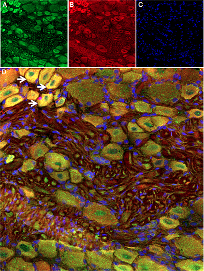
Expression of DkTx-ATTO Fluor-647N in Rat DRG Frozen Sections.
A) Anti-TRPV1 (VR1) (extracellular) Antibody (#ACC-029) was applied, 1:50, for 60 minutes at 37˚C, followed by staining with goat anti-rabbit AlexaFluor-488, resulting in green fluorescence predominantly in DRG cells. B) The same frozen sections underwent incubation with 0.4 µM of DkTx-ATTO Fluor-647N (#STD-010-FRN) for 45 minutes at 37˚C, resulting in red fluorescence in DRG cells. C) Cell nuclei were counterstained with Hoechst 33342, emitting blue fluorescence. D) Co-localization of VR1 and DkTx in DRG cells (indicated by arrows) demonstrates that DkTx serves as a reliable biomarker for VR1 channel.