Toxin-Based Protocols
Toxin-Based Live-Cell Flow Cytometry Using Conjugated Toxins
Live-cell flow cytometry with fluorophore- or biotin-conjugated venom-derived peptide toxins enables quantitative analysis of membrane proteins in living cells. These highly specific probes allow precise detection of cell surface targets while preserving cell viability. This protocol describes live-cell staining with conjugated toxins to ensure optimal detection and accurate flow cytometric or FACS analysis.
Materials
- Conjugated toxin of choice (fluorophore- or biotin-labeled).
- 1x Phosphate Buffered Saline (PBS) (02-023-1A, Sartorius).
- Streptavidin-labeled fluorophore (S21374, Invitrogen) for biotin-conjugated toxins.
Procedure
Step 1 – Reagent Preparation
1.1 Dilute the conjugated toxin according to its Certificate of Analysis.
1.2 For biotin-labeled toxins, prepare the streptavidin-labeled fluorophore according to the manufacturer’s instructions.
- Always keep fluorescent reagents protected from light during preparation and staining.
Step 2 – Preparation of Cells
2.1 Transfer 1×10⁶ cells into an Eppendorf tube.
- Centrifuge for 5 minutes at 200 g.
2.2 Discard the supernatant and wash the cell pellet by resuspending in 1 mL PBS with gentle pipetting.
- Centrifuge for 5 minutes at 200 g.
2.3 Discard the supernatant.
- Minimize shear stress to preserve cell viability and staining integrity.
Step 3 – Toxin Incubation
This step enables the conjugated toxin to selectively bind to its molecular target on the cell.
3.1 Add the toxin solution to the cells and gently resuspend by pipetting.
- Optimize toxin concentration and incubation time based on the specific target and cell type.
- Incubate 1 hour at 37°C in a humidified chamber.
Step 4 – Streptavidin (for biotin-conjugated toxins only)
A streptavidin-labeled fluorophore amplifies the signal from biotinylated toxins.
4.1 Wash cells three times with PBS: wash the cell pellet by resuspending in 1 mL PBS with gentle pipetting.
- Centrifuge for 5 minutes at 200 g.
4.2 Apply the streptavidin-conjugated fluorophore.
- Incubate 15-30 minutes at 37°C.
Step 5 – Washing
This step reduces background fluorescence and removes both unbound streptavidin and excess conjugated toxin.
- Centrifuge for 5 minutes at 200 g.
5.1 Discard the supernatant and resuspend the cells in 1 mL PBS.
- Centrifuge for 5 minutes at 200 g.
5.2 Discard the supernatant, resuspend the cells in 1 mL PBS and filter them appropriately before the FACS analysis.
Troubleshooting
| Problem | Possible Cause | Solution |
|---|---|---|
| Low signal | Fluorophore degradation, poor target expression, or inappropriate toxin concentration | Verify storage and reconstitution of toxins; confirm target protein expression in the chosen cell line; optimize toxin concentration |
| High background | Excess toxin or insufficient washing | Reduce toxin concentration and/or incubation time; increase washing steps |
| Photobleaching | Prolonged light exposure | Minimize light exposure; keep stained cells in the dark until analysis |
| Inaccurate signal interpretation | Lack of proper controls | Include negative controls: unstained cells, unlabeled toxin, and streptavidin-only (for biotin-conjugated toxins) |
Example Data
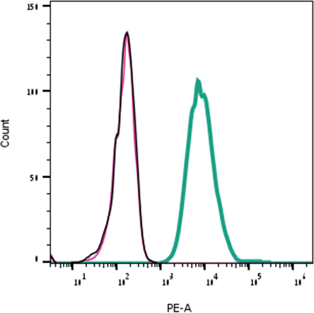
Direct flow cytometry of α-Bungarotoxin in live intact rat PC-12 cells.
___ PC-12 cells.
___ PC-12 cells + 0.1 µM α-Bungarotoxin (#B-100).
___ PC-12 cells + 0.1 µM α-Bungarotoxin-ATTO Fluor-550 (#B-100-AY).