Toxin-Based Protocols
Toxin-Based Live-Cell Imaging Using Conjugated Toxins
Conjugated venom-derived peptide toxins serve as precise probes for live-cell imaging and biodetection. These conjugates enable selective visualization of ion channels, receptors, and other membrane targets in living cells, providing key insights into their localization and dynamics. The protocol is optimized to achieve robust and reproducible data, providing consistent performance and reliable quality in live-cell imaging applications.
Materials
- Conjugated toxin of choice (fluorophore- or biotin-labeled).
- 1x Phosphate Buffered Saline (PBS) (02-023-1A, Sartorius).
- Streptavidin-labeled fluorophore (S21374, Invitrogen) for biotin-conjugated toxins.
Procedure
Step 1 – Reagent Preparation
1.1 Dilute the conjugated toxin according to its Certificate of Analysis.
1.2 For biotin-labeled toxins, prepare the streptavidin-labeled fluorophore according to the manufacturer’s instructions.
- Always keep fluorescent reagents protected from light during preparation and staining.
Step 2 – Toxin Incubation
This step enables the conjugated toxin to selectively bind to its molecular target in the cell.
2.1 Add the toxin solution to adherent cells in imaging dishes.
- Optimize the toxin concentration and incubation time based on the specific target and cell type.
- Incubate 10-60 minutes at 37°C in a humidified chamber.
Step 3 – Streptavidin (for biotin-conjugated toxins only)
A streptavidin-labeled fluorophore amplifies the signal from biotinylated toxins.
3.1 Wash the cells three times with PBS.
3.2 Apply the streptavidin-conjugated fluorophore.
- Incubate 15-30 minutes at 37°C.
Step 4 – Washing and Imaging
This step removes unbound streptavidin, excess conjugated toxin, and reduces background fluorescence.
4.1 Wash the cells three times: gently add PBS to the culture dish, swirl carefully, and remove the solution without detaching the cells.
4.2 Image using a live-cell–optimized microscope.
Troubleshooting
| Problem | Possible Cause | Solution |
|---|---|---|
| Low signal | Fluorophore degradation, poor target expression, or inappropriate toxin concentration | Verify storage and reconstitution of toxin; confirm target protein expression in the chosen cell line/tissue; optimize toxin concentration |
| High background | Excess toxin or insufficient washing | Reduce toxin concentration and/or incubation time; increase washing steps |
| Photobleaching | Prolonged light exposure | Minimize light exposure; keep stained cells/tissue in the dark until analysis |
| Inaccurate signal interpretation | Lack of proper controls | Include negative controls: unstained cells/tissue, unlabeled toxin, and streptavidin-only (for biotin-conjugated toxins) |
Example Data
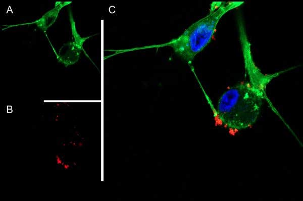
Live cell imaging of DkTx-ATTO Fluor-647N in cultured DRG cells.
Dorsal root ganglion (DRG) cells were isolated from the lumbar and thoracic regions of 3-week-old Sprague-Dawley rats and cultured with 100 ng/ml Native mouse NGF 2.5S protein (99%) (#N-240). A) CellMask™ Actin 1X solution was applied, producing green fluorescence to visualize cellular membrane. B) The same cells were then incubated with 0.1 µM of DkTx-ATTO Fluor-647N (#STD-010-FRN), resulting in red fluorescence. C) Merged live images revealed the distribution of DkTx among DRG cells. Cell nuclei were counterstained with Hoechst 33342 (blue).