Nav1.8 expression confirmed
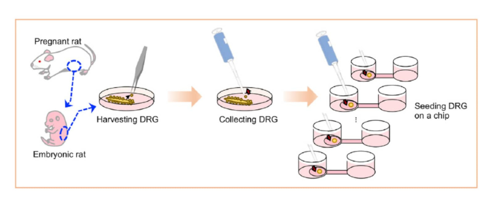
A team from Japan just published a new 3D organotypic model of peripheral sensory nerves, and it’s impressive (1). Made from rat dorsal root ganglion (DRG) explants, this model comes closer to mimicking the real peripheral nervous system than anything out there right now. We’re talking myelinated A fibers, unmyelinated C fibers, and nodes of Ranvier – all the essential structures often missing from other models. It’s the kind of thing that could change how we study nerve damage and pain.
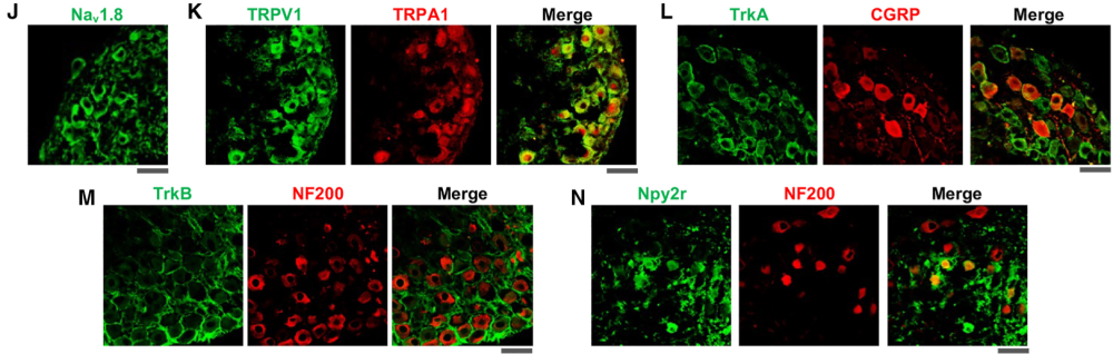
Of course, to make sure this model is the real deal, they’ve used a lot of immunohistochemistry in validation. These ex vivo neurons are packed with markers you’d see in real rat DRG: Nav1.8, TRPA1, TRPV1, TrkA, TrkB, and Npy2r. They’re all there, in ganglion-like structures, exactly like you’d expect them to be in vivo (Figure 1). The team used our Guinea pig Anti-NaV1.8 (SCN10A) Antibody (#ASC-016-GP) to confirm those NaV 1.8 channels, which are critical for nociceptive signal transmission, are right where they should be. This matters a lot when you’re trying to understand how pain signals travel – or how to stop them.
A model like this gives more labs the chance to study peripheral neuropathies, which have traditionally been hard to recreate in vitro due to the complex interactions between myelinated Schwann cells and sensory neurons. It allows for controlled, detailed examination of these interactions, disease progression, and even possible treatments. The ex vivo nerves fire under stimuli, calcium surges through, and everything behaves just like the real thing. It’s not perfect, but it’s pretty close. For anyone trying to figure out neuropathy, or how to fix it, this is a solid step forward.


 Antibody")