A technical guide to ICC and IHC
Immunocytochemistry (ICC) and immunohistochemistry (IHC) with ion channels require attention to detail. These proteins are membrane-bound, structurally complex, and often closely related to other family members. Antibody-based detection must be precise, validated, and sensitive to fixation and permeabilization conditions. This guide outlines the key technical considerations for working with membrane proteins, especially ion channels, using Alomone Labs antibodies.
Antibody Specificity and Validation
Epitope Selection
Ion channels typically share conserved domains – particularly across subtypes. Selecting antibodies that recognize unique extracellular or intracellular epitopes is critical to avoid off-target staining. Sequence alignment tools can help identify divergence between isoforms. Be sure to use antibodies with confirmed specificity data, ideally supported by peptide blocking and knockout validation.
Blocking Peptides
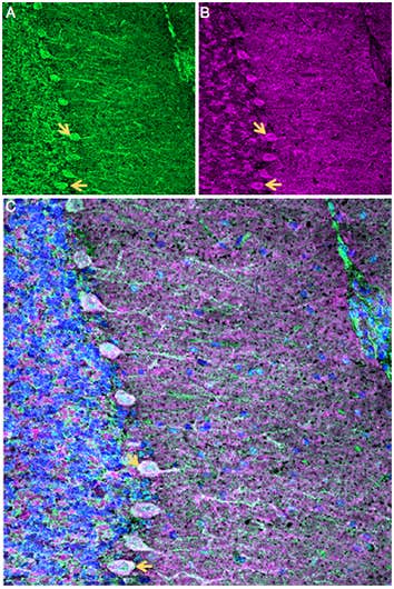
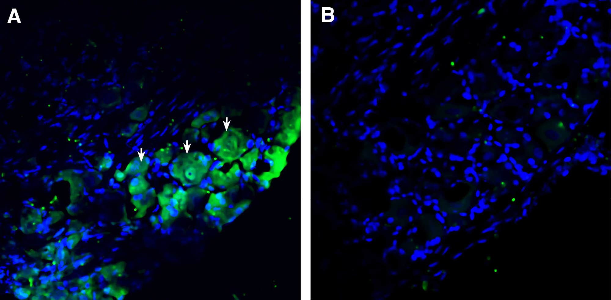
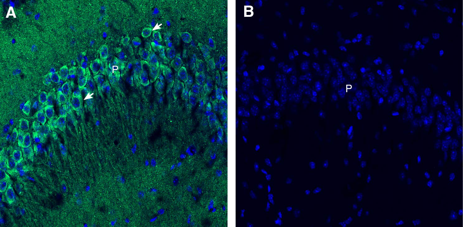
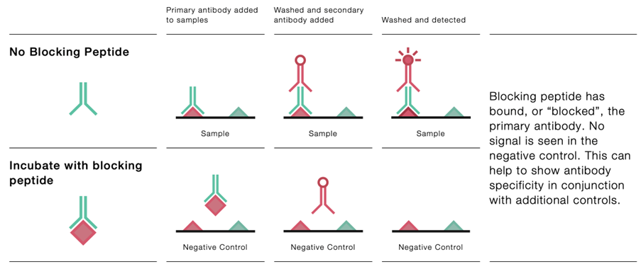
Pre-incubating the antibody with its corresponding immunogenic peptide should eliminate non-specific staining. If it doesn’t, your signal isn’t specific. All Alomone Labs antibodies raised against defined peptide sequences have a matching blocking peptide for this exact purpose.
Sample Preparation and Fixation
Fixation Protocols
Fixation preserves structure but can obscure epitopes. Paraformaldehyde (PFA) is common, but even low concentrations (2–4%) can mask delicate membrane-associated epitopes. For ion channels, especially those with short extracellular loops, over-fixation is a fast way to lose signal.
Methanol or mixed protocols (PFA followed by MeOH) may preserve antigenicity better – empirical testing is recommended.
Antigen Retrieval
If PFA fixation is required, antigen retrieval can help. Heat-induced epitope retrieval (HIER) in citrate buffer (pH 6) or Tris-EDTA (pH 9) may restore access to buried sites. But it’s not universally helpful – some epitopes degrade under retrieval conditions. Always test both retrieval and non-retrieval conditions.
Permeabilization and Blocking
Permeabilization
For intracellular targets, Triton X-100 or Tween-20 are standard. For membrane proteins, especially ion channels, use low detergent concentrations (≤0.1%). High detergent levels can extract or denature your target. Digitonin is milder and preserves membrane structure – consider it when working with delicate epitopes.
Blocking
To reduce background, use 5% normal serum (species matched to secondary antibody) or 1–5% BSA. Avoid harsh blocking agents or commercial mixes that haven’t been tested with your antibodies – some contain azides or detergents that can interfere with staining in certain assays. We have a range of carrier-free antibody options if you need an antibody free from BSA and sodium azide.
Live-Cell Labeling Considerations
Surface Epitope Accessibility
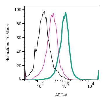
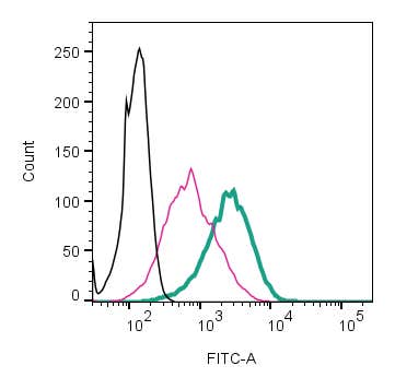
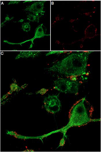
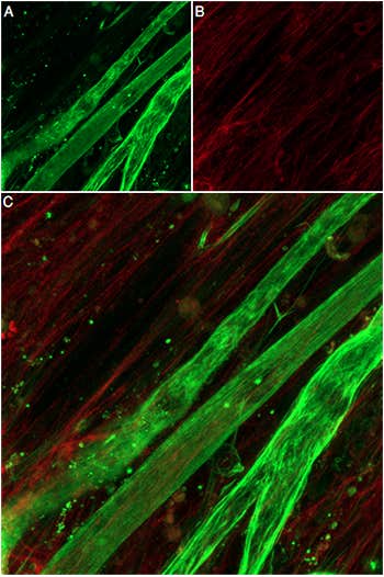
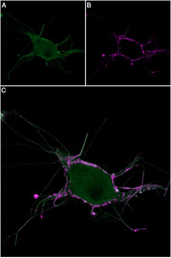
Live-cell ICC only works if your antibody targets an extracellular domain. Confirm that the epitope is externally exposed and accessible under native conditions. Do not permeabilize during live-cell work – this will compromise membrane integrity and kill your cells. To help you with this, we’ve developed a whole range of cell surface-binding antibodies that specifically bind extracellular epitopes (extracellular domain). These are ideal for tracking live cell dynamics in membrane proteins. We even have an ICC protocol specially tailored for live cells.
Temporal Dynamics
Live-cell labeling allows tracking of dynamic processes like ion channel trafficking, insertion, and internalization. Use time-lapse imaging to monitor these processes. Validate that labeling does not alter channel behavior by running paired control experiments.
Fluorophore Selection
Use bright, photostable fluorophores suitable for live-cell imaging. Directly conjugated primary antibodies (e.g., Anti-TRPV1-ATTO 488) eliminate the need for secondaries, reduce protocol time, and minimize non-specific binding. Always verify that conjugation preserves binding.
Controls and Quantification
Controls
No antibody, no signal. Omit the primary antibody for negative controls. Use tissue or cells with known expression levels for positive controls. For quantitative studies, normalize fluorescence to area, cell count, or total protein. Always include technical and biological replicates.
Blocking Peptides Again
Yes, again. Peptide pre-absorption remains one of the most reliable tests for getting a handle on specificity. If your signal disappears with a blocking peptide, that’s good. If it doesn’t, you’ve got a problem.
Quantification
Use consistent acquisition settings across samples. Don’t compare intensities from slides imaged with different exposure times or gain settings. Use software with background subtraction, intensity thresholding, and region-of-interest (ROI) tools. ImageJ and Imaris are popular choices.
Advanced Applications: 3D Tissue Clearing and Optical Imaging
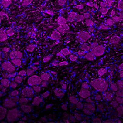
Standard sectioning limits visualization to 2D slices. For deeper imaging, use solvent-based clearing techniques like iDISCO or CUBIC. These make tissues transparent and allow antibody penetration for full-volume 3D imaging. Studies using Alomone Labs antibodies with these techniques (e.g., Anti-TRPV1, Anti-HCN4, Anti-NKCC1) show robust labeling throughout cleared brain and heart tissue.
Clearing methods must be matched with compatible antibodies (read more about it in our blog: Clearing methods). Organic solvent-based methods (e.g., iDISCO, uDISCO) often require primary antibodies with high affinity and stability. We offer validated antibodies and fluorophore-conjugated options suited for these workflows.
Functional Modulation: Agonists, Antagonists, and Blockers
For validation and functional assays, use small molecules to modulate ion channel activity. Agonists confirm functional expression. Blockers confirm specificity. We have a comprehensive set of validated ion channel modulators, including popular targets such as:
- TRPV1: Capsaicin (agonist), Capsazepine (antagonist)
- HCN channels: ZD7288 (blocker)
- NaV channels: Tetrodotoxin (TTX, blocker)
Pair these with antibody labeling to correlate structural expression with functional state.
Summary
Immunostaining of ion channels requires careful control of specificity, fixation, permeabilization, and labeling conditions. Whether you’re working with fixed or live cells, or even cleared tissues for 3D imaging, success depends on epitope accessibility, antibody validation, and experimental control. Alomone Labs antibodies are developed and tested under these exact constraints – saving you time and delivering confidence in your results.
Need help picking the right antibody or setting up your protocol? Our scientists are happy to help.