What if an antidepressant could work within hours instead of weeks?
Ketamine, a drug originally developed as a dissociative anesthetic, has transformed antidepressant research through its ability to rapidly alleviate depressive symptoms, even in some patients with treatment-resistant depression. Unlike conventional antidepressants, which typically require weeks to produce clinical benefits, ketamine can act within hours. Although its effects have traditionally been linked to NMDA receptor antagonism, the molecular mechanisms responsible for its rapid action and the persistence of its antidepressant effects remain incompletely understood.
For years, studies of ketamine-induced antidepressant effects have focused on the upregulation of brain-derived neurotrophic factor (BDNF) and the subsequent activation of the receptor TrkB. In fact, if you delete BDNF in mice, the ketamine-induced rapid mood-lifting effects vanish (1), whereas if you increase the expression of BDNF-induced extracellular signal-regulated kinase (ERK), ketamine-induced behavioral effects are enhanced (2).
However, receptors rarely act alone at the synapse, and in a study published in Science Advances, the authors examined the role of the G protein-coupled receptor (GPCR) metabotropic glutamate receptor 5 (mGluR5) – already known to be involved with ketamine’s actions (3).
Ketamine Function Requires TrkB and mGluR5
When the team blocked either TrkB or mGluR5 in mice, ketamine-induced antidepressant-related behavioral effects disappeared. What was interesting was that the two receptors seemed to be tied together via the same molecular crosstalk previously described for BDNF.
In hippocampal slices, 30 minutes of ketamine exposure drove a ~50% increase in the hippocampal synaptic CA3-CA1 field excitatory postsynaptic potential (fEPSP) slope after an hour. The TrkB antagonist ANA-12 blocked this to a similar degree as the mGluR5 negative allosteric modulator, MPEP. This same pattern was observed when examining structural plasticity, where ketamine drove a 20–30% increase in cultured hippocampal neuron spine density, but that increase was completely negated by either drug. Behaviorally, deleting mGluR5 specifically from CA1 excitatory neurons abolished ketamine-induced antidepressant-like effects, mirroring earlier work with TrkB knockouts in CA1 neurons (1). Finally, using a tool that selectively blocks TrkB-mediated noncanonical signaling (the “GBAi” tool), the team confirmed that crosstalk between the two receptors, TrkB and mGluR5, was required for ketamine to exert its antidepressant-like effects.
What Does This Crosstalk Look Like?
Probably not what you expect! There seems to be two modes of crosstalk at work. In the first mode, ketamine doesn’t activate TrkB directly; up to 100 µM of ketamine didn’t produce any calcium or ERK responses through the receptor (alone or with mGluR5). Instead, over 30 minutes, ketamine increased TrkB’s HEK cell-surface levels by 20–30%, driven by increased forward trafficking from within the cell (blocked by brefeldin A). This same increase in TrkB at the cell surface was also seen in primary hippocampal neurons, but here the increase was 40% and beyond. And with more TrkB on the cell surface, there was more ketamine-triggered BDNF caught by all of these additional receptors, and this is the crosstalk that drives potentiation.
In the second mode, once BDNF activates TrkB, mGluR5 is internalized, and also relocalized with TrkB to the dendritic spines as seen in the assays using Alomone’s Anti-mGluR5 (extracellular) Antibody (#AGC-007) (Figure 1). Fewer mGluR5s on the cell surface results in less capacity for the long-term depression (LTD) that mGluR5 usually supports. In vivo, a single ketamine injection abolished this depression in slices prepared an hour later and the effect disappeared entirely in TrkB knockout mice. Together, BDNF activation of TrkB appears to reduce the ‘brake’ effect of mGluR5 signaling by internalizing those receptors, effectively reducing the available mGluR5 pool.
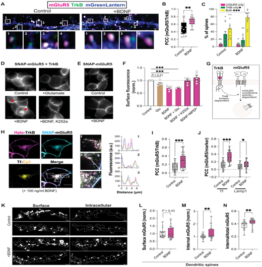
Figure 1. TrkB activation promotes mGluR5 relocalization and internalization. (A) Confocal images showing endogenous mGluR5 (red) and TrkB (green) in hippocampal neurons transduced with mGreenLantern (blue) with or without BDNF (100 ng/ml; 30 min; 37°C). Insets show dendritic spines. (B) Summary graph showing the BDNF-induced increase in TrkB/mGluR5 colocalization, as measured via Pearson’s correlation coefficient (PCC). (C) Summary graph showing an increase in the proportion of spines containing both receptors following BDNF treatment. (D and E) Representative images of HEK 293 cells showing mGluR5 internalization following treatment with BDNF only in the presence of TrkB and in the absence of the TrkB inhibitor K252a. (F) Summary graph of mGluR5 surface levels following 30 min of treatment with ligands [1 mM Glu, BDNF (50 ng/ml), 200 nM K252a, and 1 μM MPEP]. (G) TrkB activation drives mGluR5 endocytosis in a kinase-dependent mechanism, which also depends on mGluR5 constitutive activity. (H) Confocal images (left), zoomed insets (middle), and line scans (right) showing colocalization of mGluR5, TrkB, and endosomal recycling complex marker Tf-Cy3 following BDNF treatment in HEK 293 cells. PCC analysis showing an increase in TrkB/mGluR5 colocalization (G) and mGluR5 colocalization with endosomal markers (H). (K) Secondary dendritic segment (gray outline) images with surface hemagglutinin (HA)-labeled mGluR5 (left) and internalized HA-labeled mGluR5 (right). (L to N) Summary of normalized mean intensity of cell surface (L), internalized (M), and the ratio of internalized to total (N) mGluR5 within dendritic spines. Individual points represent individual cells pooled from multiple biological replicates [(B), (C), (H), and (I)], coverslips from separate biological replicates (F), or secondary dendrites from distinct neurons [(K), (L), (M) and (N)]. Unpaired t-test [(B), (I), (L), (M), and (N)], one-way ANOVA (F), or two-way ANOVA [(C), (J)] with Šidák’s multiple comparisons. All data shown as the mean ± SEM; *P < 0.05, **P < 0.01, and ***P < 0.001. Scale bars, 1 μm [(A) and (K)] or 10 μm [(D), (E), and (H)].
Figure and legend adapted from Arefin et al. (2026). 10.1126/sciadv.aec1444.
A two-mode model like this can be tested by increasing mGluR5 levels, which should also boost the effects of ketamine. To test this, the team co-administered the mGluR5 positive allosteric modulator, VU-29, with a low dose of ketamine (10mg/kg) known to not produce antidepressant-like effects. Interestingly, co-administration of VU-29 with a low dose of ketamine (10mg/kg) did produce classic antidepressant-like effects. Alone, VU-29 had no effects on behavior or synaptic strength – it only had an effect once ketamine had already engaged the pathway.
Taken together, the data reveals how ketamine drives BDNF release and boosts surface levels of TrkB, which in turn drives two modes of crosstalk with mGluR5:
- Mode 1: “signaling crosstalk” allows activation of downstream effectors to promote synaptic potentiation and spine growth. This could drive the acute antidepressant effects of ketamine over the timescale of minutes to hours.
- Mode 2: “trafficking crosstalk” pulls mGluR5 from the cell surface to reduce synaptic depression processes, aiding the synaptic strengthening effect of ketamine by lessening its reversal. This mode of crosstalk might be at the heart of ketamine-induced long-lasting antidepressant-like effects (over hours and days).
Both modes of crosstalk may, in principle, be boosted by more forward trafficking of TrkB, which in turn may provide a larger and more stable pool of receptors to drive both synergistic signaling and subsequent endocytosis.
Ketamine Function Requires Crosstalk
Despite the research team’s discussion of the therapeutic action of ketamine, it’s worth keeping in mind that the work here is from mice and cultured cells. However, this research does at least provide a redefined mechanism for an mGluR5-directed drug combined with a low dose of ketamine to extend the antidepressant window of efficacy.
But the most interesting finding here is that the BDNF receptor TrkB, and the glutamate receptor mGluR5, share the workload coming from ketamine-induced signaling. TrkB moves to the cell surface to drive synaptic potentiation, while TrkB activation pulls mGluR5 from the surface of the cell to block LTD that would otherwise reverse the ketamine effects. All in all, antidepressant durability seems to rely a lot on cutting the brakes.