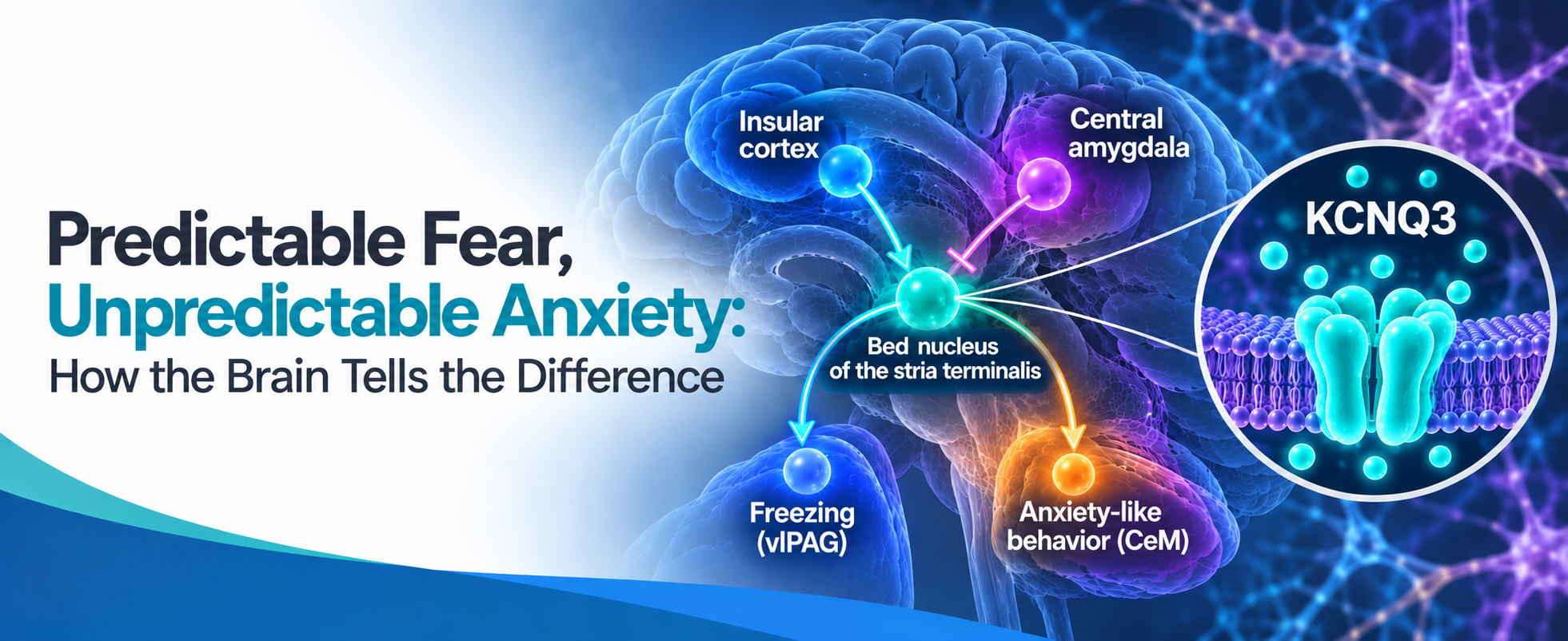
Why do unpredictable threats provoke anxiety while known dangers trigger more immediate fear? New research published in Cell Reports, reveals how the brain distinguishes between the two. Scientists identified a coordinated network involving the ventral bed nucleus of the stria terminalis (vBNST), the insular cortex (IC), and the amygdala that translates uncertain threats into freezing and anxiety-like behavior. The study also highlights the potassium channel KCNQ3 as a potential therapeutic target, offering a promising new direction for treating anxiety disorders caused by unpredictable stress.
Anxiety and fear are both products of a threat signal. Anxiety is apprehension about a threat that cannot be placed in time. Even though the danger may be plausible, nothing in the environment indicates when it will arrive, so the defensive response runs continuously rather than on cue. On the other hand, fear is the time-locked version of the same process, in which case there’s a discrete signal and a discrete response. Both are adaptive, and an animal capable of neither doesn't last long in the wild. What separates them is how much information the threat signal carries about its own timing.
Diffuse cues and unpredictable environmental events are treated as contributors to anxiety disorders, which makes the circuitry that distinguishes predictable from unpredictable threats worth identifying. The vBNST has been linked to defensive responses under diffuse, sustained, or unpredictable threat, whereas central amygdala circuits are well established in cue-evoked freezing. Freezing is the defensive immobility a rodent adopts after detecting threat, where all movement besides breathing stops. The specific cell types and mechanisms in the vBNST which support that role, and what determines their selectivity, have not been established.
Modeling Anxiety and Fear
Pavlovian fear conditioning (FC) is the assay primarily used to help understand fear and anxiety in animal models. This typically involves a neutral cue, the conditioned stimulus (CS), paired with an aversive unconditioned stimulus (US), usually a brief footshock. After a few pairings, the CS alone elicits a defensive response, which is usually freezing in mice. How much the animal learns about timing depends on the pairing: a short cue that reliably co-terminates with the shock is temporally informative, whereas a cue that is sustained, or never paired, turns up in a threatening context without indicating when danger will occur. Two cues can therefore produce the same amount of freezing while carrying quite different information about when a shock will arrive.
In recent work, Teng and colleagues present both cue types to the same animals during conditioning (1). The predictable CS (CS+) is a tone that co-terminates with a footshock, while the unpredictable CS (CS−) is an acoustically distinguishable tone, delivered pseudo-randomly and never paired. Both produce freezing after conditioning, and freezing was comparable across the two test sessions. Therefore, any neural difference cannot be explained simply by how much the animal froze, which is consistent with sensitivity to threat predictability.
A vBNST GABAergic Population Responds Selectively to the Unpaired Cue
Fiber photometry from the vBNST showed that the calcium signal in response to the CS− grew across conditioning and stayed elevated at the test. The CS+ signal did not generate a response at any point as it remained flat through habituation, flat through conditioning, and flat at the test. Restricting the recording to GABAergic neurons in Vgat-Cre mice reproduced the selectivity. The complementary Vgat− population responded to neither cue, and c-Fos counts in GAD67+ neurons agreed. Photometry revealed population activity, establishing a differential population signal rather than the behavior of individual cells across the two cues.
The authors then used optogenetic silencing to test whether that signal was required for the measured freezing response. Silencing vBNST GABAergic neurons reduced freezing to the CS− and left CS+ freezing intact, while silencing the Vgat− population changed neither.
Bulk Activation Exposes Heterogeneity Within the Population
Silencing the full GABAergic population supported the photometry results. Activating the same population produced feeding, which was unexpected. This discrepancy showed that vBNST GABAergic neurons are functionally heterogeneous, so the authors moved from cell-type-wide manipulation to ensembles defined by what activated them.
Using an activity-dependent tagging system, they labelled feeding-responsive and CS−-responsive cells separately and showed that these populations are largely distinct. Activating the CS−-responsive ensemble in a novel context, with no cue present at all, raised freezing and reduced exploration in both the open field and the Elevated Plus Maze (EPM). The total distance travelled was unchanged and feeding was unaffected, so this is a defensive phenotype rather than a general change in how much the animal moves or eats.
Shared Excitation and Cue-selective Inhibition
Isolating a CS−-responsive ensemble established which vBNST cells could generate the behavior but said nothing about why those cells respond to CS− and not CS+. Thus, the authors traced and tested their inputs. Retrograde tracing from the CS− ensemble pointed to the IC and the central amygdala as preferential sources, a pattern distinct from the inputs to the feeding ensemble.
vBNST-projecting IC neurons increased activity during both tests, and whole-cell recording confirmed a monosynaptic glutamatergic connection onto vBNST GABAergic neurons. Nonetheless, optogenetic inhibition of IC terminals in the vBNST only reduced freezing to the CS−.
These results indicated that differential IC recruitment cannot explain the selective vBNST signal. Therefore, the authors searched for inhibition recruited specifically by the CS+. vBNST-projecting lateral nucleus of the amygdala (CeL) neurons, which are roughly 80% somatostatin+, showed the opposite activity profile, with elevated calcium levels during the CS+ test but no change during the CS− test. Their terminals evoked bicuculline-sensitive inhibitory post-synaptic currents (IPSCs) in vBNST GABAergic neurons. Moreover, optogenetically activating those terminals reduced CS− freezing without affecting CS+ freezing nor producing any freezing in naive mice.
While an intriguing model here is forming, there’s still a problem, since inhibiting the CeL somtostatin neurons (CeLSOM) to vBNST projection had no effect on freezing in response to either cue. If cue-selective inhibition were the sole determinant of CS+ responsiveness, removing it should have revealed a vBNST-dependent response, and it didn't, at least not at the level of freezing output. CS+ freezing appears instead to be carried by a parallel CeLSOM to ventral lateral periaqueductal gray (vlPAG) projection. The inhibition onto the vBNST is demonstrated physiologically and is sufficient to suppress CS− freezing when activated. Whether it’s necessary to discriminate between cues isn’t established by these data.
Separable Outputs Associate With Separable Defensive Measures
The input experiments offered a putative explanation for cue selectivity, but they left a second question open, i.e., how does one activity-defined ensemble produce both freezing and anxiety-like measures?
Anterograde tracing from the CS− ensemble returned both the vlPAG and the medial central amygdala (CeM), and terminal stimulation separated the measures. Activating vBNST to vlPAG projection increased freezing without changing open-arm behavior. Activating vBNST to CeM projection reduced the animals’ exploration in the open field and EPM without changing total distance or freezing. These are activation experiments, so they associate freezing and anxiety-like measures with separable vBNST outputs rather than showing that either projection is required under normal conditions.
Chronic Unpredictable Stress Elicits the CeM-associated Phenotype
Thus far, the mechanism revealed an acute defensive circuit responding to cues within a session. The more interesting question is whether repeated unpredictable stress leaves the same ensemble persistently overactive, which would connect cue processing to something lasting.
A week of chronic unpredictable stress, with randomized stressors several times daily, raised spontaneous activity in CS−-responsive vBNST cells recorded in the home cage, alongside increased anxiety-like behavior. Silencing those cells, or the vBNST to CeM projection, reversed the behavioral phenotype. Silencing vBNST to vlPAG projections did not. CeL SOM activity was unchanged after stress, which argues against altered activity in that inhibitory input, though IC to vBNST activity was never measured after stress. Therefore, the relative contributions of altered afferent drive and altered intrinsic excitability remain unresolved.
KCNQ3 and Intrinsic Excitability
Chronic stress increased spontaneous activity in the tagged ensemble, but the circuit recordings could not distinguish whether that came from altered input or from changes in the cells themselves. Thus, the authors screened transcripts from recently activated vBNST neurons, then tested membrane excitability directly.
The screen returned 60 genes under the ion channel activity term and 12 of them were potassium channels. Kcnq3 mRNA levels showed the largest decrease, as shown by qRT-PCR. Immunofluorescence with Alomone’s Anti-KCNQ3 Antibody (#APC-051) confirmed lower KCNQ3 levels in GAD67+ vBNST neurons after the CS− test compared to the CS+ test (Figure 1). Western blotting with the same antibody showed reduced KCNQ3 protein levels after chronic stress.
In slice recordings from CS−-responsive neurons after stress, the intrinsic excitability was increased and the M-current relaxation was reduced. Restoring KCNQ3 by overexpression in those cells prevented the rise in home-cage calcium activity and reduced anxiety-like behavior. Local microinjection of Alomone’s Retigabine (#R-100), a KCNQ2-5 opener with relatively high potency at KCNQ3-containing channels, reduced both the electrophysiological and the behavioral abnormalities.
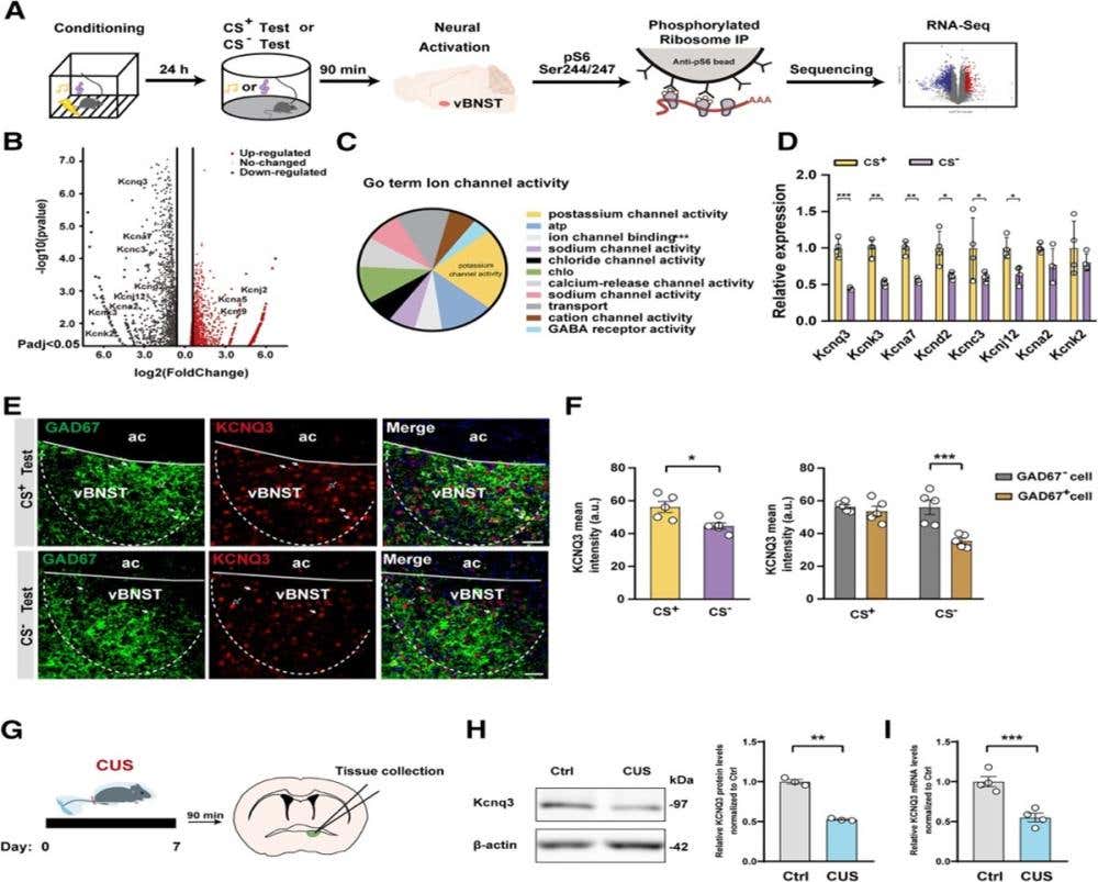
Figure 1. A reduction in KCNQ3 levels was identified following a CS− test and CUS. (A) Schematic of the pTRAP-Seq experiment design and the behavioral paradigm. (B) Differential expression of pTRAP-Seq genes between CS− and CS+ tests. (C) GO analysis of ion channel activity as overrepresented in the CS− test group (GO: 0005216), p+ channels. (D) The expression of the reduced K+ family genes was verified by qRT‒PCR (n = 4 animals per group). (E) Representative images of GAD67+ (green) and KCNQ3+ (red) immunofluorescence in the vBNST. Solid arrows indicate neurons co-expressing GAD67 and KCNQ3, and hollow arrows indicate neurons expressing KCNQ3 alone. Scale bar, 50 μm. (F) Quantification of KCNQ3 immunofluorescence intensity (n = 5 animals per group). (G) Schematic of the vBNST tissue collection experiment design and the behavioral paradigm. (H) Representative expression of KCNQ3 by Western blots in control (Ctrl) and CUS samples. The migration of MW markers is shown on the right side of the Western blot. Quantification of the Western blots (n = 3 animals per group). (I) The CUS group showed a significant reduction in Kcnq3 mRNA levels (n = 4 animals per group). ∗p < 0.05, ∗∗p < 0.005, ∗∗∗p < 0.001. Data are presented as the mean ± SEM.
Figure and legend adapted from Teng et al. (2025). https://www.sciencedirect.com/science/article/pii/S2211124724014979.
What Survives the Limitations
The circuit claim holds up within the limits of the design: coordinated IC excitation and CeLSOM inhibition, a vBNST ensemble whose activity tracks the unpaired cue, and two output projections whose activation dissociates freezing from anxiety-like measures.
The molecular claim is narrower than it first appears. KCNQ3 was also reduced in feeding-responsive cells. Thus, the change appears to follow recent activation across at least the two ensembles tested here rather than marking unpredictable threat specifically. KCNQ3 regulates the excitability of these neurons; it doesn't, based on these data, distinguish the event that recruited them. What the study supports is that KCNQ3 functions as a regulator of excitability in stress-recruited vBNST neurons. Neither that result nor the cue design establishes a molecular or neuronal code specific to unpredictability. Either way, the data remain a great step forward in our understanding of the neuronal circuitry in anxiety.