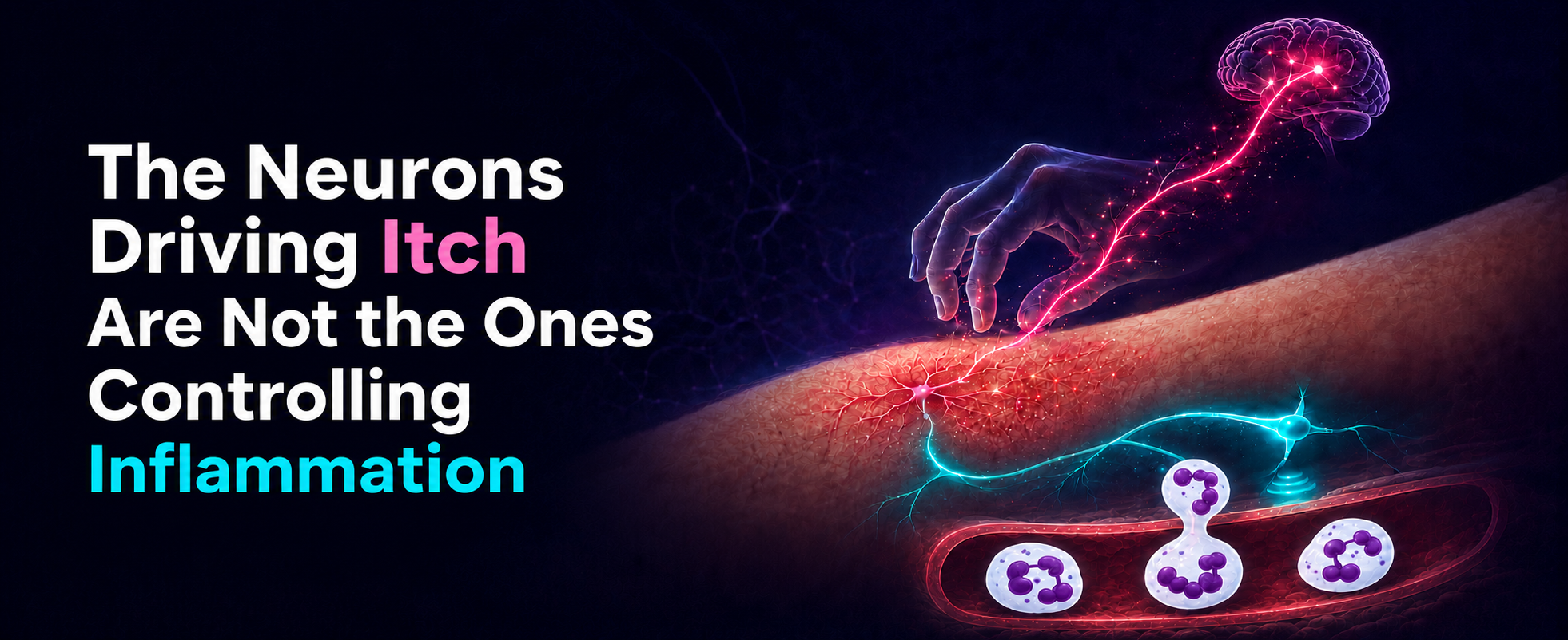
In allergic contact dermatitis, itch and skin inflammation usually get lumped together as two faces of one problem: the skin is inflamed and itchy, the person scratches it, the scratching makes it worse. The obvious assumption is that it’s the same sensory nerves feeding both parts of the problem, right? Well, in a study published in Immunity, Voisin et al. (1) pulled this problem apart and found there are two distinct nociceptor populations, each with their own job and neither one is covering for the other when the other is gone. Remove the wrong neurons, and the scratching goes down, but the inflammation gets worse. Remove a different population, and the itch disappears without the same inflammatory response.
Global Nociceptor Depletion Produced Opposite Phenotypes
The team started with a broad depletion experiment. In NaV1.8-Cre;iDTA mice, which lack all nociceptor subsets, 1,4-dinitrofluorobenzene (DNFB) induced contact hypersensitivity (CHS), which produced more severe skin pathology, including increased neutrophils and monocytes in the skin by day 4. But the same animals showed less itch-evoked scratching across the model.
Thus, nociceptors were doing two opposing things at once, where some were helping drive the itch response, while others were limiting the inflammatory reaction. This is likely why this problem has been hard to resolve. If you ablate or silence nociceptors as a bulk population, you collapse separate circuits into one readout. The net phenotype depends on which function dominates in that model, time point, tissue site, and inflammatory context. It’s a great approach for proving sensory neurons matter but less useful for figuring out which neurons do what!
Going Finer
The finer view came from retrograde tracing plus single-cell RNA-seq analysis. The team labelled skin-projecting dorsal root ganglion (DRG) neurons with DiI, a fluorescent cell membrane dye, sorted the traced cells, and sequenced them. One population stood out: NP1 neurons, a specific subclass of nonpeptidergic, small-diameter, unmyelinated C-fiber sensory neurons characterized by the expression of Mas-related G-protein-coupled receptor member D (MrgprD).
They later validated CHS-associated changes in neuronal marker expression by immunostaining for markers using antibodies directed against IB4, GFRα2 and TH, as well as Alomone’s Anti-P2X3 Receptor (extracellular) Antibody (#APR-026) for the non-peptidergic lineage and Alomone’s Anti-TRPV1 (VR1) Antibody (#ACC-030) for the peptidergic side.
When they inflamed the skin and looked again, the MrgprD+ NP1 population had changed in what they seemed to be doing. These neurons switched on a whole panel of regeneration-associated genes normally seen after nerve injury, such as Atf3, Sprr1a, and Gal (all confirmed by qPCR, not just transcriptomics), even though no nerve was cut. It seemed inflammation alone was enough to push NP1 neurons into a transient, injury-like regenerative state. “Reprogrammed” was the word they used, and it fits, since the cells adopt a distinct molecular identity and then, once things settle, revert.
Which Population Does What
To ask whether the reprogrammed NP1 neurons carry the itch, they selectively ablated them. GINIP-hDTR mice were crossed onto NaV1.8-Cre mice to hit the NP1/C-low-threshold mechanoreceptors (LTMR) compartment. In a separate experiment, an Atf3-CreERT2;iDTA mice were generated to target specifically the neurons that had switched on the injury program. Take those cells out and scratching is strongly reduced or abolished, while the neutrophilic inflammatory response is not worsened. Thus, the reprogrammed NP1 population is primarily an itch driver, rather than the brake on neutrophil-heavy inflammation.
The other arm of the experiments required a different tool. To deplete the TRPV1+ peptidergic neurons in vivo, they administered Alomone’s Resiniferatoxin (#R-400) subcutaneously over three consecutive days with escalating doses of 30, 70, then 100 µg/kg. This toxin ablated the TRPV1+ nociceptors rather than just desensitize them. As a result, they saw inflammation become markedly worse, with a sharp rise in neutrophil infiltration and tissue damage (Figure 1). But the itch was untouched.
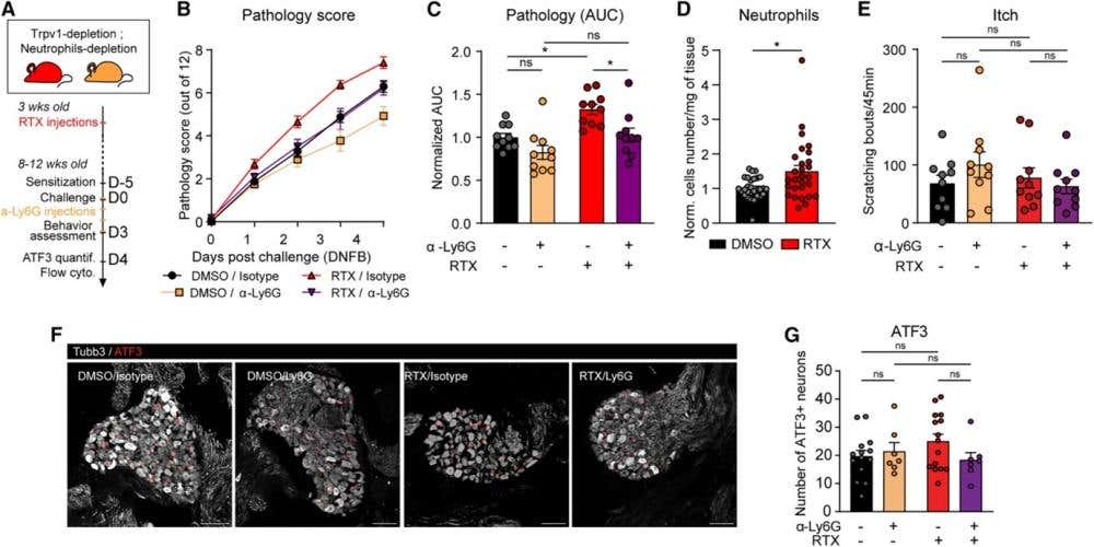
Figure 1. In CHS, TRPV1+ neurons regulate inflammation, but not itch. (A) Schematic diagram of the experimental plan. (B and C) Time course (B) and associated area under the curve (AUC) (C) of the skin pathology score induced by DNFB in DMSO/isotype (black), DMSO/anti-Ly6G (yellow), RTX/isotype (red), and RTX/anti-Ly6G (purple) mice (n = 10 per group, two independent experiments). (D) Number of neutrophils normalized (Norm.) per experiment in skin from DMSO or RTX mice (n = 28–29 per group, three independent experiments). (E) Scratching bouts at day 3 in DMSO/Isotype (black), DMSO/anti-Ly6G (yellow), RTX/isotype (red) and RTX/anti-Ly6G (purple) mice (n = 10 per group, two independent experiments). (F and G) Representative images of DRG sections [beta-tubulin (white) and ATF3 (red)] (F) and associated number of ATF3+ neurons per DRG section per mouse (G) in DMSO/isotype (black), DMSO/anti-Ly6G (yellow), RTX/isotype (red), and RTX/anti-Ly6G (purple) at day 4 (n = 10 per group, two independent experiments). Scale bar, 100 μm. Values represent the mean ± SEM. (B) ∗p < 0.05, ∗∗p < 0.01, ∗∗∗∗p < 0.0001, Mann-Whitney U-test, (C, D, and G) ∗p < 0.05, two-way ANOVA with Sidak’s post test, ns, not significant.
Figure and legend adapted from Voisin et al. (2026). https://doi.org/10.1016/j.immuni.2026.03.020.
The results of these experiments revealed two circuits with opposite functions: (1) reprogrammed MrgprD+ NP1 neurons regulate the itch – remove them and the itch goes away while inflammation remains; and (2) TRPV1+ peptidergic neurons regulate (restrain) neutrophilic inflammation – remove these and inflammation increases while itch remains. And neither compensates for the other.
The Mechanism That Isn't There Yet
The obvious next question is how the TRPV1+ neurons hold the neutrophils back. It’s a logical question, and here the paper is honest about its limits. The intuitive answer is the neuropeptides, since TRPV1+ neurons are loaded with neuropeptides like CGRP, substance P, and PACAP, all with known immune-modulating activity. To test this, they blocked them individually: BIBN4096 was used to inhibit CGRP signaling, PACAP6-38 was used to repress PACAP, and an anti-SP antibody was used to suppress substance P. All of the treatments exhibited no major effects on the pathology. Whatever the TRPV1+ circuit is using to keep neutrophils in check, it doesn't appear to run through these three neuropeptides, at least not any one of them alone.
They did manage to deduce that neutrophils are the effectors: depleting them with anti-Ly6G in Resiniferatoxin (RTX)-treated animals reversed the disease exacerbation without touching itch nor the NP1 injury response. Even though they figured out the consequence, the upstream signal from the TRPV1+ neuron to the neutrophil remains unknown.
A New Look for Nociceptors
This research neatly reframed what nociceptors are doing in inflamed skin. There’s no single pro-scratch, pro-pathology route, but a division of labor where one population protects the tissue while another generates the symptom. That's a more useful model for understanding the biology and it came directly from resolving the population rather than treating “nociceptors” as one thing.
Also, since itch and inflammation are on separable circuits, new treatments might be able to quiet one without disabling the other. Silence the reprogrammed NP1 itch driver and leave the TRPV1+ brake on neutrophils intact. That's a genuinely different targeting approach from anything that hits all sensory neurons at once.
The authors also note that the mechanism remains unresolved, and that the pharmacological blockade experiments should be backed up with knockout models before pinning too much relevance on the negative peptide results. Farthermore, the central question of how the signal from a TRPV1⁺ neuron tells a neutrophil to stay out is still unclear since the peptide candidates came up empty. Therefore, the interesting answer of this story is the part nobody's written yet.