Normalizing failure and encouraging better documentation.
You followed the protocol. You ran all the right controls. You used fresh reagents, calibrated equipment, and stuck to the script. Last week’s western blot was clean. Today? Blank.
Every researcher hits this wall. The “But I did everything right!” moment is more a rite of passage than an anomaly. And when it happens, it’s tempting to assume you’ve messed something up. Or that the antibody’s bad. Or maybe you’re just not cut out for this.
None of that is true.
In reality, most experimental failures come not from big mistakes, but from small, undocumented variables. Tiny changes – pipetting order, buffer composition, exact timing – can unravel everything. And unless you’ve logged those details, you’ll be left chasing ghosts in your protocol.
Reproducibility Depends on What You Remember to Write Down
You might think you’ll remember how long you incubated that gel. You won’t. You might assume the buffer you made last week was the same as the one today. It probably isn’t.
Most PhD students (and plenty of postdocs) underestimate how much procedural nuance sits in their heads, not their notebooks. But memory degrades fast, especially when you’re juggling multiple experiments, meetings, and deadlines. What felt like a small deviation at the time becomes an invisible source of variability weeks later. That’s how reproducibility quietly fails.
But this isn’t about competence. If your lab notebook can’t reconstruct your exact steps, you’ve lost the ability to learn from the result.
Failure Happens. Documentation Gives It Meaning
It’s easy to take a failed experiment as a reflection of your ability. If you’re early into your research years, it’s easy to internalize these collapses as personal. You’ll hear senior researchers say, “You must have messed something up” or “Did you even read the protocol?” Neither helps. Because science breaks more often than it works. What matters is whether you can figure out why. And that depends entirely on what you’ve recorded.
Take that failed western blot. If your notes just say, “Standard protocol,” that’s a dead end. Which version? What were the actual incubation times? Was it a new lot of antibody? Was the blocking solution fresh or reused? Was the shaker set to the same speed?
If these details are missing, then all of a sudden you’re guessing rather than troubleshooting.
Write Things Down to Protect Your Sanity
Detailed notes serve the experiment and, more importantly, they support you. When the data fall apart, the protocol fails, and you’ve got three deadlines looming, your notebook is the only thing that can help you make sense of it all.
Memory is limited. Documentation isn’t. When you’ve tracked your buffers, timing, order of steps, and instrument settings, you can actually learn from what broke. Without that, all you’ve got is the feeling that something went wrong.
The Details That Most People Miss
Even the most seasoned researcher can and will forget to note things down. When you’ve run that IHC 300 times already, you stake a lot for granted. Don’t do that. Take the time to note the important things down. Start with the variables that derail reproducibility the most:
- Pipetting sequence
- Reagent lot and expiration
- Buffer prep and pH
- Room temperature and humidity
- Actual – not intended – incubation times
- Calibration state of instruments
- Well layout and sample order
Small shifts here can produce large changes in output. The more precisely you record them, the more likely it is that your results can be explained and replicated.
Choose Tools That Help You Rule Things Out
When something fails, the first question should be, “What changed?” Sometimes the answer to that might be a new antibody against the same target. So, look there. Not all suppliers validate antibodies and other reagents in the same way. And if validation was consistent, then this question stays open.
At Alomone Labs we make sure each lot of reagents is validated in multiple assays. Antibodies for example might have knockout, IHC, and western blot validation data, along with blocking peptide controls readily available. The protocols and datasheets have validation data, results from work published by other, independent scientists, and a host of open data (like antibody immunogen sequences). This narrows the field of uncertainty.
When you’re troubleshooting, knowing the reagent works and has been validated removes one variable. That lets you focus on what actually went wrong, rather than chasing the wrong culprit.
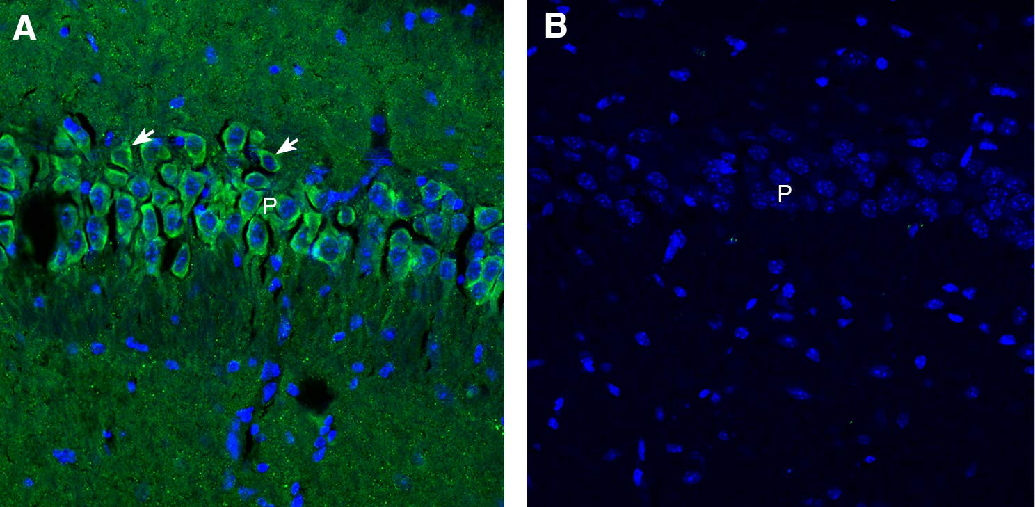
Ever tried to troubleshoot an experiment weeks later and couldn’t remember what went wrong? That’s where negative controls like rabbit IgG isotype (Figure 1) and blocking peptides (Figure 2) come in. They don’t just validate your results in the moment – they give you a clear baseline to refer back to.
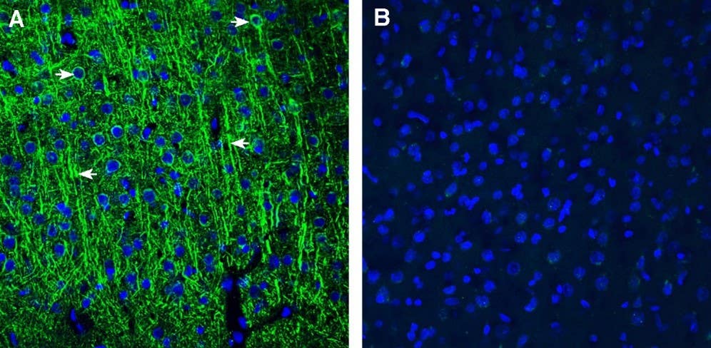
Figure 1. Comparison of immunostaining with SSTR1 compared to immunostaining with isotype control antibody in rat cortex. A) Immunohistochemical staining of perfusion-fixed frozen rat brain sections with Anti-Somatostatin Receptor Type 1 (extracellular) Antibody (#ASR-001) (1:300), followed by goat anti-rabbit-AlexaFluor-488. SSTR1 immunoreactivity (green), appears in neuron outlines, in both dendrites (right pointing arrows) and soma (left pointing arrows). B) Staining of sequential sections with Rabbit IgG Isotype Control (#RIC-001) (1:300), followed by goat anti-rabbit-AlexaFluor-488, shows background signal. Cell nuclei are stained with DAPI (blue). [Image sourced from alomone.com].
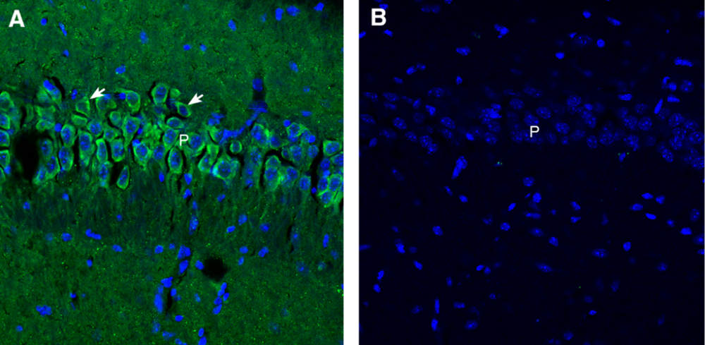
Figure 2. Comparison of Synaptojanin-1 immunostaining with and without blocking peptide in mouse hippocampal CA1 region. A) Immunofluorescent staining of free-floating frozen brain sections from a paraformaldehyde-perfused mouse using Anti-Synaptojanin 1 Antibody (#APZ-061) (1:300), followed by goat anti-rabbit-Alexa Fluor® 488. Synaptojanin-1 immunoreactivity (green) is evident in neuronal profiles within the CA1 region (arrows). B) Pre-incubation of the primary antibody with Synaptojanin 1 Blocking Peptide (#BLP-PZ061) abolishes the staining signal, confirming specificity. Nuclei are counterstained with DAPI (blue). P = pyramidal layer. [Image sourced from alomone.com].
When the Right Reagent Tells the Right Story
In the lab, clear notes keep you from chasing ghosts – the same goes for your reagents. At Alomone Labs, each antibody lot is validated by western blot. Additional validation – such as IHC, blocking peptide controls, and reference to knockout assay data from external studies – is performed when relevant. The result: you start with certainty, backed by rigorous validation. Our datasheets back it up with real validation images, independent citations, and open details like immunogen sequences. We also provide detailed protocols on our website to guide you step-by-step, making sure you get the best out of each product. With more than 30,000 published studies citing our products, you can see the evidence for yourself in our Citation Tool. If the data takes a turn, having proven reagents on your side means you can pinpoint the real variable at play.
Repeatability Starts with What You Write and What You Use
You don’t have to write everything down forever, but you do need to record the right things with enough detail that you can return to the method and understand what you did. Over time, that record becomes the most valuable tool in your workflow.
We can’t control your notebook. But our reagents are designed to minimize doubt – and that matters when the signal disappears and your supervisor is waiting for answers.
You shouldn’t always see failure in the lab as a sign that something’s broken. Rather, it’s a sign that something needs to be found. The best way to find it is by knowing what happened the first time: clearly, exactly, and on paper.
Useful links