
Antibody-based detection remains central to characterizing neurological disease pathology. These protein-level readouts offer cell-type resolution, subcellular specificity, and compatibility with tissue and fluid samples. While some markers track core pathological events – like tau phosphorylation in Alzheimer’s or α-synuclein accumulation in Parkinson’s – others reveal secondary processes such as gliosis, immune infiltration, or synaptic remodeling.
Here we outline the most commonly used antibody-detectable protein markers across major neurological diseases and highlight their detection platforms.
Alzheimer’s Disease (AD)
Amyloid Precursor Protein (APP), Amyloid-β (Aβ) peptides
Proteolytic products of APP and Aβ are involved in plaque formation; Aβ accumulation is central to AD pathology.
Phosphorylated Tau (p-tau)
Tau is normally a microtubule-associated protein, but hyperphosphorylation causes it to misfold and aggregate into neurofibrillary tangles. Tau is a key pathological marker that is detected in brain tissue and cerebrospinal fluid (CSF) samples.
Neurofilament Light Chain (NfL)
NfL is a sensitive marker of axonal damage and neurodegeneration that is elevated in the CSF and blood of AD patients. NfL expression correlates with AD severity and progression. It is a non-specific neurodegeneration marker (also elevated in other diseases such as ALS, MS, etc.), but it’s highly useful for tracking disease activity.
Glial Fibrillary Acidic Protein (GFAP)
GFAP is an astrocyte intermediate filament protein, upregulated in reactive astrocytosis. It indicates gliosis and neuroinflammation in AD.
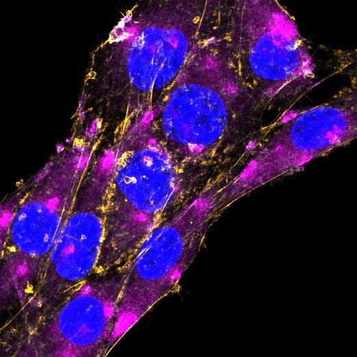
Figure 1. Expression of GFAP in rat C6 glioma cells. Fixed and permeabilized cells were stained with Anti-GFAP-ATTO Fluor-647N Antibody (#AFP-001-FRN), (1:200, magenta) and with CellMask Orange Actin Tracking Stain (orange). The cell nuclei were stained with Hoechst 33342 (blue).
Triggering Receptor Expressed on Myeloid Cells 2 (TREM2)
TREM2 is a microglial receptor involved in the immune response; TREM2 mutations are linked to an increased AD risk.
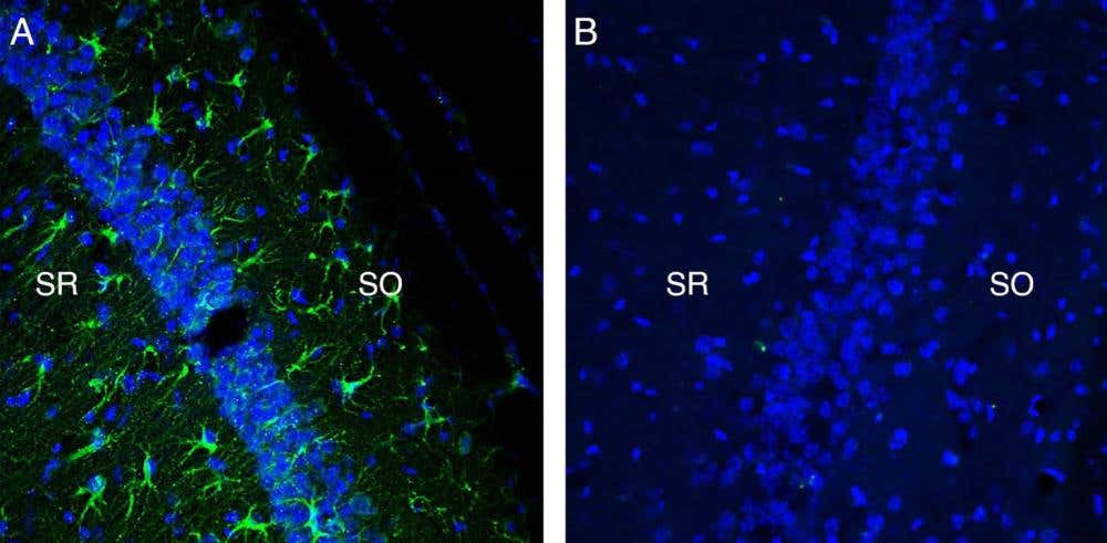
Figure 2. Hippocampal TREM2 expression in the kainic acid–induced excitotoxicity model. Immunohistochemical staining of perfusion-fixed frozen mouse brain sections collected 4 days after kainate injection with Anti-TREM2 (extracellular) Antibody (#ANR-018), (1:300), followed by goat anti-rabbit-AlexaFluor-488 antibody. A) Staining in the mouse hippocampal CA1 region showed TREM2 immunoreactivity (green) in astrocytes located in the stratum oriens (SO) and the stratum radiatum (SR) of the hippocampus. B) Pre-incubation of the antibody with TREM2 (extracellular) Blocking Peptide (#BLP-NR018), suppressed staining. The cell nuclei were stained with DAPI (blue).
Parkinson’s Disease (PD)
α-Synuclein
α-Synuclein is a presynaptic protein normally involved in synaptic vesicle regulation. In PD it misfolds and aggregates into Lewy bodies and Lewy neurites – key PD pathological events.
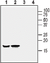
Figure 3. Western blot analysis of rat (lanes 1 and 3) and mouse (lanes 2 and 4) brain lysates: 1,2. Anti-Alpha-Synuclein Antibody (#APZ-035), (1:400). 3,4. Anti- α-Synuclein Antibody, pre-incubated with Alpha-Synuclein Blocking Peptide (#BLP-PZ035).
Dopamine Transporter (DAT)
DAT is responsible for regulating dopamine reuptake from the synaptic cleft into presynaptic terminals; DAT loss correlates with nigrostriatal degeneration in PD.
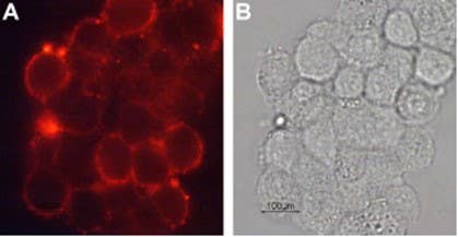
Figure 4. Expression of DAT in rat pheochromocytoma PC12 cells. Cell surface detection of DAT in live intact rat PC12 cells. A) Cells stained with Anti-Dopamine Transporter (DAT) (extracellular) Antibody (#AMT-003), (1:100), followed by goat anti-rabbit- AlexaFluor-594 antibody (red). B) Live brightfield image of the same cells.
DJ-1 [a.k.a. Parkinson Disease Protein 7 (PARK7)]
DJ-1 is involved in the oxidative stress response. Mutations in DJ-1 cause rare, early-onset, autosomal recessive PD.
Parkin (a.k.a . PARK2)
Parkin is a E3 ubiquitin ligase involved in mitochondrial quality control; mutations in parkin cause autosomal recessive PD.
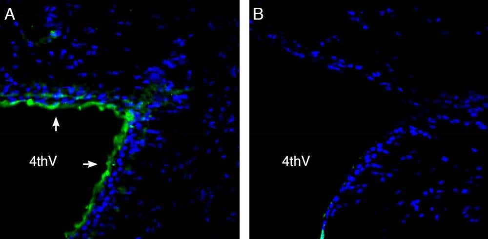
Figure 5. Expression of parkin in the rat brain 4th ventricle. Immunohistochemical staining of perfusion-fixed frozen rat brain sections with Anti-Parkin Antibody (#ANR-103), (1:200), followed by goat anti-rabbit-AlexaFluor-488 antibody. A) Parkin immunoreactivity (green) appears along the wall of the 4th cerebral ventricle (arrows). B) Pre-incubation of the antibody with Parkin Blocking Peptide (#BLP-NR103), abolishes staining. The cell nuclei were stained with DAPI (blue). 4th V= 4th ventricle.
Amyotrophic Lateral Sclerosis (ALS)
TAR DNA-Binding Protein 43 (TDP-43)
TDP-43 is an RNA/DNA-binding protein involved in RNA metabolism. In most ALS cases, TDP-43 becomes mislocalized from the nucleus to the cytoplasm, where it forms toxic aggregates that contribute to motor neuron degeneration.
Neurofilament Light Chain (NfL)
NfL is an axonal injury marker elevated in both the CSF and serum of ALS patients. NfL is associated with ALS disease progression.
Synaptic Vesicle Proteins [Vesicle-Associated Membrane Protein 2 (VAMP2), Synaptosome Associated Protein 25 (SNAP-25)]
VAMP2 and SNAP-25 are Involved in neurotransmitter release, and their altered expression reflects synaptic dysfunction in ALS.
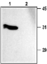
Figure 6. Western Blot analysis of rat brain membranes. 1. Anti-SNAP-25 Antibody (#ANR-001), (1:1000). 2. Anti-SNAP-25 Antibody, pre-incubated with SNAP-25 Blocking Peptide (#BLP-NR001).
Epilepsy
Glial Fibrillary Acidic Protein (GFAP)
GFAP is associated with astrocyte activation and gliosis found in epileptic brain tissue.
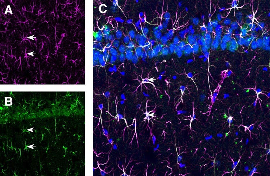
Figure 7. Expression of GFAP and Thyroid-Stimulating Hormone (TSH) Receptor in the rat hippocampus CA1 region. Immunohistochemical staining of perfusion-fixed frozen rat brain sections with Anti-GFAP-ATTO Fluor-647N Antibody (#AFP-001-FRN), (1:80) and Anti-TSH Receptor (TSHR) (extracellular)-ATTO Fluor-488 Antibody (#ATR-006-AG), (1:80). A) GFAP immunoreactivity (magenta) appears in astrocytes (arrows). B) TSH receptor immunoreactivity (green) appears in astrocytic processes (arrows). C) Merging of the two images confirms specific astrocytic staining. The cell nuclei were stained with DAPI (blue).
S100 Calcium-Binding Protein 3 (S100B)
S100B is a calcium-binding protein expressed by astrocytes, which is elevated in brain injury and epilepsy.
Neuronal Specific Enolase (NSE)
NSE is a glycolytic enzyme expressed in neurons. It is a neuronal injury marker, and elevated levels have been reported in the CSF and serum after seizures, traumatic brain injury, and ischemia.
Multiple Sclerosis (MS)
Myelin Basic Protein (MBP)
MBP is a major myelin component; increased CSF levels of MBP indicate demyelination in MS.
Glial Fibrillary Acidic Protein (GFAP)
GFAP is a marker of astrogliosis that is associated with central nervous system (CNS) inflammation in MS.
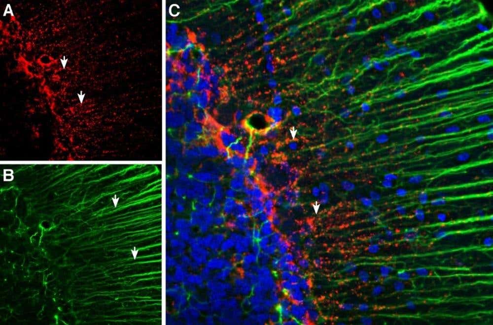
Figure 8. Multiplex staining of Connexin-43 (CNX-43) and GFAP in the rat cerebellum. Immunohistochemical staining of perfusion-fixed frozen rat brain sections with Guinea pig Anti-Connexin-43 Antibody (#ACC-201-GP), (1:200), followed by goat anti-guinea pig-AlexaFluor-594 antibody (panel A) and Anti-GFAP Antibody (#AFP-001), (1:1200), followed by goat anti-rabbit-AlexaFluor-488 antibody (panel B). A) CNX-43 immunoreactivity (red) appears as positive puncta (arrows). B) GFAP immunoreactivity (green) appears along the Bergmann glial processes (arrows point at examples). C) Merging of the two images shows CNX-43 puncta distributed along the Bergmann glial processes. The cell nuclei were stained with DAPI (blue).
Cluster of Differentiation 3, 4, 8 (CD3, CD4, CD8)
CD3, CD4, and CD8 are T-cell markers that indicate immune cell infiltration in MS lesions.
Oligoclonal Bands [(OCBs) Immunoglobulins]
OCBs in the CSF are the most established diagnostic biomarker for MS. They signify intrathecal antibody synthesis, indicating a sustained immune response in the CNS.
Huntington’s Disease (HD)
Mutant Huntingtin Protein (mHTT)
mHTT is a mutant huntingtin protein, which contains an expanded polyglutamine tract that induces neuronal toxicity, particularly in the striatum and cortex.
Neurofilament Light Chain (NfL)
NfL is a non-specific marker of axonal injury, consistently elevated in the CSF and blood in HD patients. Its levels correlate with disease onset, severity, and progression.
Synaptic Proteins [Synaptophysin (SYP), Postsynaptic Density Protein 95 (PSD95)]
Synaptic proteins such as SYP (presynaptic marker) and PSD95 (postsynaptic density protein) show altered expression and distribution in HD brains and animal models. Their reduction correlates with synaptic degeneration and impaired neurotransmission in HD pathology.
- Guinea Pig Anti-Synaptophysin Antibody (#ANR-013-GP)
- Anti-Synaptophysin Antibody (#ANR-013)
- Anti-PSD-95 Antibody (#APZ-009)
- Guinea Pig Anti-PSD-95 Antibody (#APZ-009-GP)
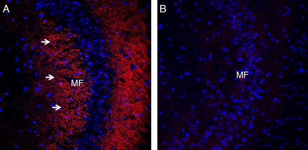
Figure 9. Expression of Synaptophysin in the mouse hippocampus. Immunohistochemical staining of perfusion-fixed frozen mouse brain sections using Guinea Pig Anti-Synaptophysin Antibody (#ANR-013-GP), (1:200), followed by goat anti-guinea pig Alexa Fluor 594 antibody. A) Staining in the hippocampal CA3 region shows immunoreactivity (red) in the mossy fiber terminal zone (MF, arrows). B) Pre-incubation of the antibody with Synaptophysin Blocking Peptide (#BLP-NR013) suppresses the staining. Cell nuclei are counterstained with DAPI (blue).
Additional Antibody-Detectable Protein Markers in Neurological Disorders
- Alzheimer’s disease: APP, Aβ, p-tau, NfL, GFAP, TREM2
- Parkinson’s disease: α-Synuclein, DAT, DJ-1, Parkin
- Amyotrophic lateral sclerosis: TDP-43, NfL, VAMP2, SNAP-25
- Epilepsy: GFAP, S100B, NSE
- Multiple sclerosis: MBP, GFAP, CD3, CD4, CD8, OCBs
- Huntington’s disease: mHTT, NfL, SYP, PSD-95
Additional Protein Markers Relevant in Neurological Disorders
Take a look at our other blog if you need a more detailed look at synaptic markers for pre- and postsynaptic regions. Otherwise, here are some of the more commonly used markers for other neurological disorders.
Synaptophysin (SYP)
SYP is a presynaptic vesicle glycoprotein widely used as a universal marker of synaptic density. SYP exhibits altered expression in AD, HD, schizophrenia, and epilepsy.
Postsynaptic Density Protein 95 (PSD-95)
PSD-95 is a major scaffolding protein expressed at excitatory synapses, critical for synaptic stability and signaling. Altered levels of PSD-95 are linked to AD, autism spectrum disorders, schizophrenia, and other synaptopathies.
Neuroligin, Neurexin Families
These are synaptic adhesion molecules essential for synapse formation and function. Neuroligin mutations are associated with autism spectrum disorders, while neurexin alterations are implicated in schizophrenia and epilepsy.
- Anti-Neuroligin 1 (extracellular) Antibody (#ANR-035)
- Anti-Neuroligin 2 (extracellular) Antibody (#ANR-036)
- Anti-Neuroligin 3 (extracellular) Antibody (#ANR-037)
- Anti-Neurexin 1α (extracellular) Antibody (#ANR-031)
- Anti-Neurexin 3α (extracellular) Antibody (#ANR-033)
- Neuroligin & Neurexin Antibody Explorer Kit (#AK-595)
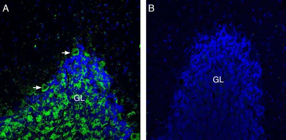
Figure 10. Expression of Neuroligin-3 in the rat cerebellum. Immunohistochemical staining of perfusion-fixed frozen rat brain sections using Anti-Neuroligin-3 (extracellular) Antibody (#ANR-037), (1:200), followed by goat anti-rabbit Alexa Fluor 488 antibody. A) Neuroligin-3 immunoreactivity (green) is observed in the granule layer (GL) and in some Purkinje cells (arrows). B) Pre-incubation of the antibody with Neuroligin-3 (extracellular) Blocking Peptide (#BLP-NR037) suppresses the staining. Cell nuclei are counterstained with DAPI (blue).
Synapsins (Synapsin I, II)
Synapsins are a family of phosphoproteins that regulate synaptic vesicle trafficking and neurotransmitter release. Altered expression and phosphorylation patterns of synapsins have been reported in epilepsy (hyperexcitability). Alterations in synapsin levels are also observed in AD (synaptic dysfunction) and mood disorders (depression, bipolar disorder), reflecting their role in neurotransmission.
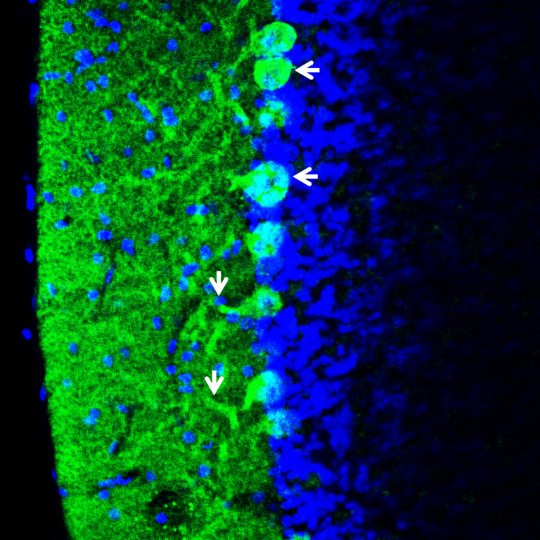
Figure 11. Expression of synapsin-1 in the mouse cerebellum. Immunohistochemical staining of a perfusion-fixed frozen mouse brain section using Anti-Synapsin I (SYN1) Antibody (#ANR-014), (1:400), followed by anti-rabbit-AlexaFlour-488 antibody. Synapsin-1 staining (green) appears in Purkinje cell bodies (horizontal arrows) and in segments of the dendritic tree (vertical arrows). The nuclei were stained with DAPI (blue).
Gephyrin
Gephyrin is a postsynaptic scaffolding protein localized at inhibitory GABAergic synapses. Loss or dysfunction of gephyrin is implicated in epilepsy, autism, and schizophrenia.
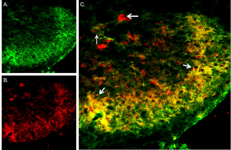
Figure 12. Multiplex staining of VGAT and Gephyrin in the rat spinal cord. Immunohistochemical staining of perfusion-fixed frozen rat spinal cord sections using Anti-Gephyrin Antibody (#AIP-005), (1:300), followed by anti-rabbit-AlexaFlour-488 antibody, and Anti-Vesicular GABA Transporter (VGAT)-ATTO Fluor-594 Antibody (#AGT-005-AR), (1:80). A) Gephyrin staining (green) appears in the superficial layer of the spinal dorsal horn, and some cellular profiles were stained. B) VGAT staining (red) appears in the superficial layer of the spinal dorsal horn and in cell profiles. C) Merged images of A and B reveal co-localization (arrows) and separate expression in some cells. The cell nuclei were counterstained with DAPI (blue).
Vesicular Glutamate Transporters 1, 2 (VGLUT1, VGLUT2), Vesicular GABA Transporter (VGAT)
VGLUT1/2 and VGAT are markers for excitatory and inhibitory presynaptic terminals, respectively. Their altered distribution or expression is found in epilepsy, autism, and neurodegeneration. These markers are widely used in immunohistochemistry and immunocytochemistry to map excitatory vs. inhibitory synapses.
- Guinea Pig Anti-VGLUT1 Antibody (#AGC-035-GP)
- Anti-VGLUT1 Antibody (#AGC-035)
- Guinea Pig Anti-VGLUT2 Antibody (#AGC-036-GP)
- Anti-VGLUT2 Antibody (#AGC-036)
- Glutamate Transporter Antibody Explorer Kit (#AK-440)
Calcium/Calmodulin-Dependent Protein Kinase II (CaMKII)
CaMKII is a synaptic enzyme critical for synaptic plasticity, long-term potentiation, and memory formation. CaMKII is dysregulated in AD and other cognitive disorders.
Shank Family Proteins (Shank1, Shank2, Shank3)
Shank family proteins are large scaffolding proteins localized to excitatory postsynaptic densities. They organize the following processes: (1) receptor clustering, (2) signaling complexes, and (3) spine morphology. Mutations in these family members are linked to autism spectrum disorders and intellectual disability.
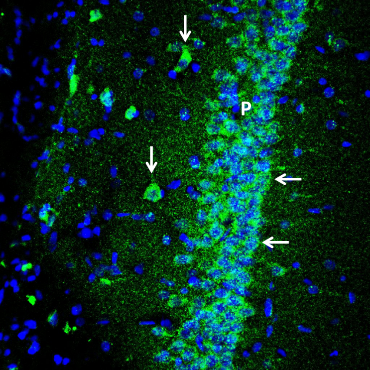
Figure 13. Expression of Shank1 in the rat hippocampus. Immunohistochemical staining of a perfusion-fixed frozen rat brain section using Anti-Shank1 Antibody (#APZ-011), (1:300), followed by goat anti-rabbit-AlexaFluor-488 antibody. Shank1 staining (green) is detected in the interneurons (vertical arrows) and in the pyramidal layer (P, horizontal arrows). The nuclei were stained with DAPI (blue).
Soluble NSF Attachment Protein Receptor (SNARE) Complex Proteins (Syntaxin-1, Synaptobrevin/VAMP2)
SNARE complex proteins are key components of the synaptic vesicle fusion machinery and are altered in neurodegenerative and psychiatric disorders.
Additional Synaptic Markers Commonly Used in Neurological Disorder Research
For a more detailed look at markers in specific regions, see our Pre- and Post-Synaptic Markers Guide.
- SYP: Presynaptic vesicles (AD, HD, epilepsy, schizophrenia)
- PSD-95: Postsynaptic density (AD, autism, schizophrenia)
- Neuroligins/Neurexins: Synaptic adhesion (autism, schizophrenia, AD)
- Synapsins: Presynaptic terminals (epilepsy, AD, mood disorders)
- Gephyrin: Inhibitory postsynaptic (epilepsy, autism, schizophrenia)
- VGLUT1, VGLUT2, VGAT: Presynaptic terminals (epilepsy, autism, neurodegeneration)
- CaMKII: Postsynaptic density (AD, cognitive disorders)
- Shank proteins: Postsynaptic density (autism spectrum disorders)
- Syntaxin-1, VAMP2: Presynaptic SNARE (AD, ALS, schizophrenia)
Together, these markers and kits provide essential tools to investigate neuronal integrity, glial activation, synaptic structure, and immune dynamics across diverse neurological disorders. Immunohistochemistry and immunocytochemistry remain the mainstay for spatial profiling, while fluid-based immunoassays extend the diagnostic potential of CSF and blood. As new antibodies improve specificity and signal-to-noise ratios, these reagents continue to refine both basic research and clinical stratification.


-ATTO Fluor-594 Antibody")


 (extracellular) Antibody")


 Antibody")
 Antibody")
 Antibody")
 Antibody")
 Antibody")
 Antibody")
 Antibody")


 (extracellular)-ATTO Fluor-488 Antibody")
