Overview
Type: Synthetic peptide
Form: Lyophilized powder
VGLUT2 Blocking Peptide (#BLP-GC036) is the original antigen used for immunization during Anti-VGLUT2 Antibody (#AGC-036) generation. The blocking peptide binds and ‘blocks’ Anti-VGLUT2 primary antibody, this makes it a good negative reagent control to help confirm antibody specificity in western blot and immunohistochemistry applications. This control is also often called a pre-adsorption control.
Applications: wb, ihc
For research purposes only. not for human use
Applications
Demonstration of Pre-adsorption control
 Western blot analysis of rat brain membranes (lanes 1 and 4), mouse brain membranes (lanes 2 and 5) and human SH-SY5Y neuroblastoma cell lysate (lanes 3 and 6):1-3. Anti-VGLUT2 Antibody (#AGC-036), (1:400).
4-6. Anti-VGLUT2 Antibody, preincubated with VGLUT2 Blocking Peptide (#BLP-GC036). Expression of VGLUT2 in rat neocortexImmunohistochemical staining of rat frozen brain sections using Anti-VGLUT2 Antibody (#AGC-036), (1:400). VGLUT2 (red) is detected in pyramidal neurons (arrow). The same section was stained with DAPI (blue).
 Multiplex staining of VGLUT2 and P2X7 Receptor in rat spinal cordImmunohistochemical staining of perfusion-fixed frozen rat spinal cord sections using Anti-VGLUT2 Antibody (#AGC-036), (1:600) and Anti-P2X7 Receptor-ATTO Fluor-550 Antibody (#APR-004-AO), (1:100). A. Vesicular glutamate transporter 2 labeling followed by goat-anti-rabbit-Alexa-488 (green). B. The same section labeled for P2X7 receptor (orange). C. Merge of A and B demonstrates partial co-localization of VGLUT2 and P2X7 receptor in dorsal horn and in lateral column (L. Col., arrow). Cell nuclei were stained with DAPI (blue).
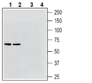 Western blot analysis of rat brain membranes (lanes 1 and 3) and mouse brain membranes (lanes 2 and 4):1-2. Guinea Pig Anti-VGLUT2 Antibody (#AGC-036-GP), (1:200).
3-4. Guinea Pig Anti-VGLUT2 Antibody, preincubated with VGLUT2 Blocking Peptide (BLP-GC036).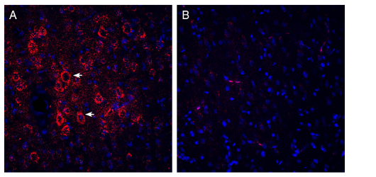 Expression of VGLUT2 in rat parietal cortex.Immunohistochemical staining of perfusion-fixed frozen rat brain sections with Guinea Pig Anti-VGLUT2 Antibody (#AGC-036-GP), (1:200), followed by goat anti-Guinea Pig-AlexaFluor-594. A. VGLUT2 immunoreactivity (red) appears in pyramidal neuronal profiles (horizontal arrows). B. Pre-incubation of the antibody with VGLUT2 Blocking Peptide (BLP-GC036), suppressed staining. Cell nuclei are stained with DAPI (blue).
Properties
Sequence
- (C)EDGKPLEVPEKK, corresponding to amino acid residues 45-56 of rat VGLUT2 (Accession Q9JI12).

Accession (Uniprot) Number Q9JI12
Peptide Confirmation Confirmed by amino acid analysis and mass spectrometry.
Purity >70%
Storage Before Reconstitution Lyophilized powder can be stored intact at room temperature for two weeks. For longer periods, it should be stored at -20°C.
Reconstitution 100 µl double distilled water (DDW).
Concentration After Reconstitution 0.4 mg/ml.
Storage After Reconstitution -20°C.
Antigen Preadsorption Control 1 µg peptide per 1 µg antibody.
Standard Quality Control Of Each Lot Western blot analysis.
