Immunohistochemistry Protocols
Immunohistochemistry (IHC) Protocols for Frozen Sections: Multiplex Staining
Target multiple proteins simultaneously with these multiplex IHC protocols using antibodies from the same or different host species.
Most immunohistochemistry (IHC) protocols describe experiments using only one antibody. In practice, however, you often need multiple antibodies against multiple targets. As the number of targets increases, so does the complexity of your multiplex experiment. However, with the right protocol, You can easily resolve the issues of managing antibodies from different host species.
Here, we describe our multiplex IHC protocol using both rabbit and guinea pig polyclonal primary antibodies on the same floating tissue sections.
If you have any problems, please see our extensive troubleshooting guides.
Materials
Buffers & Solutions
Select ONE lysis buffer depending on the method used:
- Lysis Buffer A (for Enriched Membrane Fraction Preparation)
○ 4 mM HEPES (pH 7.4)
○ 320 mM Sucrose
○ 5 mM EDTA (pH 8)
○ EDTA-free Protease Inhibitor Cocktail (11836170001, cOmplete™, Roche) - Lysis Buffer B (for Brain Synaptosome/P2 Preparation)
○ 1 mM Phenylmethylsulfonyl fluoride (PMSF)
○ Lysis Buffer A - Lysis Buffer C (for Total Tissue Lysate Preparation)
○ 50 mM Tris (pH 7.4)
○ 1% Triton X-100
○ 5 mM EDTA (pH 8)
○ EDTA-free Protease Inhibitor Cocktail (11836170001, cOmplete™, Roche)
The following buffer is required only when performing synaptosome extraction:
- Extraction Buffer
○ 50 mM Tris (pH 9.0)
○ 150 mM NaCl
○ 1% NP-40
○ 1 mM PMSF
○ 0.5% Sodium deoxycholate
○ EDTA-free Protease Inhibitor Cocktail (11836170001, cOmplete™, Roche)
Other
- Liquid nitrogen
- Dounce homogenizer
- High-speed motorized tissue homogenizer
- Ice bucket
- Heating block or water bath
- Rocking platform
- Tube rotator
- Refrigerated centrifuge (700 × g, 37,000 × g)
- Ultracentrifuge (100,000 × g)
- Scalpel or sterile razor blade
- Pre-chilled microcentrifuge tubes
Preparation and Antigen Retrieval
- Rinse the floating sections with IHC phosphate-buffered saline (IHC-PBS) for 2 x 5 minutes.
IHC-PBS (pH 7.4) Reagent Concentration Volume/Weight Na2HPO4 0.2 M 80 ml NaH2PO4 0.2 M 16 ml NaCl 8 g Double distilled water 860 ml - For antigen retrieval and to quench endogenous peroxidase activity, incubate the floating sections with IHC-PBS with 0.2% hydrogen peroxide, 0.2% Triton X-100*, and 20% methanol, for 25 minutes at room temperature.
- Rinse the sections with IHC-PBS for 2 x 5 minutes.
*If your primary antibody targets an extracellular protein, reduce the Triton X-100 to 0.05% in both the primary and secondary antibody solutions.
Two options for multiplex IHC are available here:
A. Primary Antibodies Raised in Different Hosts
B. Primary Antibodies Raised in the Same Host
OPTION A: Primary Antibodies Raised in Different Hosts
- Incubate the sections with a cocktail containing two primary antibodies:
a. The first raised in rabbit (1:200 to 1:400)
b. The second raised in guinea pig (1:200 to 1:400)
Dilute both antibodies in Multiplex Antibody Solution for 1 hour at room temperature.
*If your primary antibody targets an extracellular protein, reduce the Triton X-100 to 0.05% in both the primary and secondary antibody solutions.Multiplex Antibody Solution Reagent % of Final Volume IHC-PBS 95.65 Triton X-100* 0.3 Tween-20 0.05 Normal goat serum (NGS) 2 Normal donkey serum 2 - Incubate the sections at 4°C overnight.
- Rinse the sections with IHC-PBS containing 2% NGS and 2% NDS for 2 x 5 minutes.
- Incubate the sections with a cocktail of diluted secondary antibodies:
a. Anti-rabbit conjugated to a fluorescent dye
b. Anti-guinea pig conjugated to a different fluorescent dye
Dilute both antibodies in Multiplex Antibody Solution for 1 hour at room temperature. - Incubate the sections at 4°C overnight.
- Proceed to Mounting and Detection.
OPTION B: Primary Antibodies Raised in the Same Host
- Incubate the sections with rabbit primary antibody (un-conjugated) diluted in Antibody Solution for 1 hour at room temperature.
*If your primary antibody targets an extracellular protein, reduce the Triton X-100 to 0.05% in both the primary and secondary antibody solutions.Antibody Solution Reagent % of Final Volume IHC-PBS 97.65 Triton X-100* 0.3 Tween-20 0.05 Normal goat serum (NGS) 2 - Incubate the sections at 4°C overnight.
- Rinse the sections with IHC-PBS containing 2% NGS for 2 x 5 minutes.
- Incubate the sections with the secondary antibody (goat anti-rabbit conjugated to a fluorescent dye) in Antibody Solution for 1 hour at room temperature.
- Incubate the sections at 4°C overnight.
- Rinse the sections with IHC-PBS containing 2% NGS for 2 x 5 minutes.
- Incubate the sections with 2% normal rabbit serum (NRS) (to saturate residual binding ability of the secondary goat anti-rabbit antibody), for 1 hour at room temperature.
- Rinse the sections with IHC-PBS containing 2% NGS for 2 x 5 minutes.
- Incubate the sections with rabbit primary antibody conjugated to a different fluorescent dye, diluted 1:50–1:60 in Antibody Solution for 1 hour at room temperature.
- Incubate the sections at 4°C overnight.
- Proceed to Mounting and Detection.
*If your primary antibody targets an extracellular protein, reduce the Triton X-100 to 0.05% in both the primary and secondary antibody solutions.
Mounting and Detection
- Rinse the sections with IHC-PBS (containing 2% NGS) for 2 x 5 minutes.
- Mount sections on slides and dry for 2 hours to overnight.
- Stain the mounted sections with DAPI stain (5 mg/ml stock solution in deionized water or dimethylformamide (DMF); dilute to 500 nM in IHC-PBS for final use) to label all cells in the field. Incubate in DAPI at room temperature for 5 minutes.
- Apply coverslips using the adhesive Immu-MountTM (ShandonTM).
- Detect with a microscope.
Troubleshooting and Tips
| Problem | Possible Cause | Solution |
|---|---|---|
| Low protein yield | Tissue not fully homogenized; insufficient buffer | Homogenize thoroughly; increase buffer-to-tissue ratio. |
| Protein degradation | Warm samples; missing protease inhibitors | Keep cold; add fresh EDTA-free inhibitors; work quickly. |
| Poor solubilization | Fibrous tissue; mild buffer insufficient | Increase homogenization time; use stronger buffer if compatible. |
| High background (mild lysis) | Partial extraction of membrane components | Increase centrifugation time/speed; carefully remove supernatant. |
| Inconsistent replicates | Variable tissue input or processing | Standardize tissue amount; keep steps consistent. |
Example Data
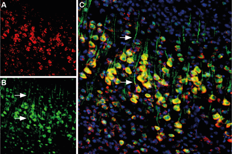
Multiplex staining of calnexin and presenilin-1 in the mouse cortex, using two rabbit primary antibodies.
The sections were incubated with a cocktail of rabbit Anti-Calnexin conjugated with ATTO red (ACS-009-AR) and rabbit Anti-Presenilin conjugated with ATTO green (AIP-011-AG) antibodies. A) Calnexin staining (red) appeared in the neuronal profiles. B) Presenilin-1 staining (green) in the same section appeared in the neuronal profiles and apical dendrites (arrows). C) A merged image of A and B demonstrated the colocalization of calnexin and presenilin-1 in several neurons (arrows). The cell nuclei are stained with DAPI (blue) as the counterstain.
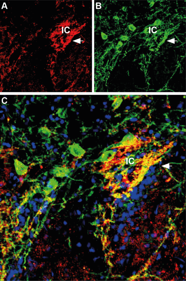
Multiplex staining of synapsin II (SYN2) and GABA(A) α1 receptor in the rat striatum using primary antibodies from different hosts.
The sections were incubated with a cocktail of rabbit Anti-Synapsin II (ANR-015) and guinea pig Anti-GABA(A) α1 receptor (AGP-083) antibodies, followed by incubation with donkey anti-rabbit Cy3 (red) and goat anti-guinea pig Alexa 488 (green). A) Synapsin II (red) appeared in the Islands of Calleja (arrow). B) The GABA(A) α1 receptor (green) was detected in the Islands of Calleja. C) A merged image of A and B demonstrated partial colocalization of synapsin II and GABA(A) α1 receptor. The cell nuclei are stained with DAPI (blue).