Mice from a pain research model used since 2001 seize under experimental stress.
The CaV2.2 knockout (KO) mouse has been a fixture of pain and anxiety research since 2001. Its appeal has always been obvious, since N-type calcium channels (1) sit at presynaptic terminals and gate neurotransmitter release; (2) carry neuropathic pain signals; and (3) are the target of ziconotide, the cone snail peptide used for otherwise chronic pain. The experimental process would typically be to delete the channel, watch the pain and anxiety phenotypes drop away, and build out the mechanism. Thousands of experiments have leaned on exactly that logic.
A new characterization study in Scientific Reports shows something the CaV2.2 KO mice users have been sitting on without knowing it: under repeated experimental testing, these mice have seizures.
Only 3 of 31 KOs briefly seized in this study but fully recovered without showing adverse events or behavior (1). But an earlier study by the same group, running more intensive testing, recorded seizures in 5 of 9 animals, 3 of them sick enough to be euthanized. The pattern the authors propose is that experimental stress, accumulated over weeks of frequent handling and testing, pushes the KO mice toward a seizure-prone state. Histology supports an early epileptogenic outcome, with reactive astrocytes in the hippocampus, activated microglia in the cortex, all without neuronal loss. The presynaptic location of CaV2.2 suggests that a KO alters the excitability of neurons and, consequently, their metabolic profile, therefore a metabolomic analysis was conducted.
Metabolomics analysis revealed downregulated palmitoylethanolamide (palmitoyl-EA, PEA) a lipid that reduces seizure frequency in epileptic models, alongside upregulated hypoxanthine and platelet-activating factor (both flagged in seizure and stroke work).
So, what does this mean for those using this mouse line? If you’ve conducted pain studies on this line, you’ve been running them on animals carrying a latent, stress-inducible seizure liability that nobody had cataloged. That’s both a confounding and a welfare problem, and it stayed invisible because the line was validated once, cross-sectionally, for the phenotype it was built to display. Nobody ran the test long enough to see the rest, which affected the reliability of the mouse model. A model is only as trustworthy as its characterization, and characterization is not a one-time event.
Every claim above depends on one fact: that the CaV2.2 channel is actually gone. If CaV2.2 is still partially expressed, the seizures, the gliosis, and the shifted metabolites could originate from something else entirely. The KO confirmation here rests on a Western blot against the α1B subunit, using Alomone’s anti-CACNA1B antibody (Anti-CACNA1B (CaV2.2) Antibody (#ACC-002)), corroborated by label-free proteomics. That single Western blot is what gives weight to every downstream difference to be attributed as a CaV2.2 effect rather than background noise. It is perhaps the least glamorous figure in the paper and the one the whole argument hangs on (Figure 1).
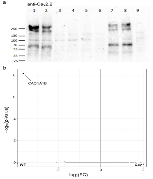
Figure 1. Absence of the α1B subunit in CaV2.2−/− mice. a) A Western blot analysis revealed the absence of the murine CaV2.2 channel (~240 kDa) in CaV2.2−/− (samples 3–6, 9) but not wild-type (WT; samples 1, 2, 7, 8) brain homogenates. The blot was cropped to size and labeled with numbers; otherwise, the original blot image remained unchanged. The original images are depicted in Supplementary Fig. 2. b) Proteomics demonstrated that the only significantly altered protein in the CaV2.2−/− vs. WT comparison is the CACNA1B protein, which is the α1B subunit of the CaV2.2 channel. A protein is considered significantly altered at p < 0.05 and a fold change (FC) < 0.
Figure and legend adapted from Wintz et al. (2026). https://doi.org/10.1038/s41598-026-60827-w.
The same logic runs through the rest of the characterization of the CaV2.2 KO mouse line. The inflammation and neuron loss results are only as good as the GFAP, Iba1 and NeuN antibodies used to study them. The metabolite shifts are only as good as the mass spectrometry identification and the subsequent database matching. Reliable models are assembled from validated reagents upward. Swap in an antibody that cross-reacts, or a marker nobody has checked in this specific tissue, and the phenotype you report may be an artifact you have mistaken for a mechanism.
This is exactly the territory where new approach methodologies (NAMs) get mentioned. Organoids, iPSC-derived neurons, and in silico systems all reduce reliance on poorly characterized whole animal models, improve reproducibility, and sidestep part of the welfare issue. For a lot of mechanistic questions about N-type channel function, that case is strong and getting stronger.
But this paper also marks the edge of it. The seizure phenotype didn’t appear in a dish – it emerged from a whole organism under weeks of accumulated experimental stress, an interaction between handling, physiology, and behavior no current in vitro system reproduces. Emergent, context-dependent behavior still requires the analysis of the animal. That does not weaken the case for better models, if anything it strengthens it. If the whole organism work is genuinely irreplaceable for these questions, then characterizing those organisms properly, longitudinally, with reagents you can trust, stops being optional.
The lesson here is not to distrust the mouse, especially since the KO remains a perfectly good tool for what it was built to do. The lesson is that model reliability must be built, not assumed. It comes from validated antibodies, honest longitudinal characterization, and a willingness to publish the results you were not looking for. Over a quarter of a century of published clean pain data, and the seizures were there the whole time. Remarkably, it’s just that someone finally ran the test long enough to find them.