For decades, the hyperpolarization-activated inward current (Ih) was the pain current neuroscientists didn't talk about. They measured it, logged it, filed it, and mostly left it at that. In sensory neurons of the dorsal root ganglion (DRG)—the small-diameter C-fibers that carry nociceptive signals to the spinal cord—the Ih was barely present; a faint inward sag when the membrane was pushed negative, with a modest amplitude and low density compared to the same current in heart or brain. The hyperpolarization-activated cyclic nucleotide-gated (HCN) channels 1 through 4, which conducted the Ih were better known for their role in cardiac pacemaking. In the context of pain, they were plausible contributors but not compelling ones. What little evidence existed was pharmacological: the blunt Ih blocker ZD7288 showed some analgesic effects in rodents, but it inhibited every HCN isoform with equal indifference.
The assumption underneath all of this was reasonable enough. The nociceptor Ih was probably what the Ih was expected to be everywhere else: a stabilizing, rhythmic, background conductance. This made it worth measuring, yet not worth pursuing. That assumption was wrong, but identifying exactly how it was wrong—which isoform, which mechanism, which pain condition—took the better part of a decade.
What the Immunostaining Revealed
In 2012, a group from Bristol published a detailed atlas of HCN1 and HCN2 protein expression in rat DRG neurons using Alomone’s rabbit polyclonal antibodies [Anti-HCN1 Antibody (#APC-056) and Anti-HCN2 Antibody (#APC-030)] (1). The antibodies had been pre-characterized against channels expressed in COS-7 or HEK cells, but the Bristol group went further, adding Western blot pre-absorption controls and siRNA knockdown of HCN2 as a specificity check before reporting a single staining pattern.
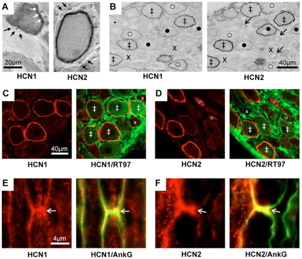
Figure 1. HCN1 and HCN2 immunostaining. A and B: ABC immunostaining. A: Utilizing the X100 objective, HCN1 and HCN2 ring staining (white arrows) was observed over the neuron perimeter and not in satellite cells (black arrows). Thus, it is likely membrane associated (sn = satellite cell nucleus). B: In adjacent sections, HCN1 and HCN2 show co-localization, with some differences in intensity or location. Symbols in B: ‡ ring staining for both HCN1 and HCN2; x neither; o clear HCN1 ring but weaker HCN2; • clear HCN2 ring but weaker HCN1. C–F: Double immunofluorescence staining in L5 DRG neurons. C and D: HCN1 (C) or HCN2 (D) ring staining (red) is present in large, myelinated, neurofilament-rich (RT97+, green) neurons. There is also cytoplasmic HCN2 staining in a few RT97−, unmyelinated, small neurons (fine arrow in D). Symbols in C and D indicate examples of staining with: ‡ both antibodies; x neither; o clear HCN2 cytoplasmic but not neurofilament staining. E and F: Representative X100 images of HCN1 (E) and HCN2 (F) (both in red) with Ankyrin G (AnkG, green) to indicate the nodes of Ranvier (arrows) in longitudinal sections of the normal L5 dorsal root nerve. Yellow is indicative of co-localization.
Figure and legend adapted from Acosta, C. et al. (2012). https://doi.org/10.1371/journal.pone.0050442.g001.
In normal animals, they found that HCN1 expression tracked with conduction velocity, which was high in large fast Aβ fibers and lower in small Aδ and C-fiber neurons. HCN2 followed the same gradient. Ring membrane immunostaining for both isoforms was largely absent in small C-fiber neurons under normal conditions, consistent with the low Ih amplitudes recorded in vivo. Thus far, this confirmed the consensus that there was not much going on in nociceptors.
However, the study had a second aim: to examine inflammation. Therefore, the group looked at what happened 1 day after complete Freund's adjuvant (CFA)-induced cutaneous inflammation. In this case, HCN2 immunostaining in small DRG neurons—the putative nociceptors—increased significantly, while HCN1 did not. By day 4, after CFA-induced cutaneous inflammation, HCN2 levels in small neurons had substantially recovered, tracking the same 1 day peak in C-nociceptor spontaneous firing neurons identified in prior work from the same group. The effect was transient, but the subtype specificity was unambiguous: HCN2, but not HCN1, was responding to the inflammatory signal in exactly the cell population that matters for pain.
The key question was whether this reflected functional significance. Protein upregulation in a fixed tissue slice is one thing, but whether more HCN2 in a nociceptor actually changes how that nociceptor fires is another. The immunostaining had established a correlation between HCN2 expression and inflammatory pain states. The next challenge was determining whether HCN2 was merely associated with nociceptor sensitization or was actually required for it.
The Genetic Proof
Around the same time, Edward Emery and colleagues at Cambridge asked that question directly and answered it in a way that made the field pay attention (2). Their experiment was built around a conditional knockout (cKO) of HCN2 deleted specifically in NaV1.8-expressing neurons—exactly the small-diameter nociceptors where protein upregulation had been observed under CFA-induced cutaneous inflammation. In these mice, the cyclic adenosine monophosphate (cAMP)-sensitive component of Ih was abolished, while the remaining Ih was cAMP-insensitive, consistent with it being carried primarily by HCN3.
On the behavioral side, naïve HCN2 knockout mice had normal acute pain thresholds. However, when inflammation was induced with prostaglandin E2 (PGE2) or carrageenan, these mice failed to develop heat hyperalgesia. That specificity ran in an unexpected direction: inflammatory mechanical hyperalgesia was abolished by neither nociceptor-specific HCN2 deletion nor by ZD7288, a selective HCN channel blocker. Nociceptors detecting heat pain and those detecting mechanical pain appear to diverge downstream of the cAMP signal. The data suggested that HCN2 was driving the thermal response while something else was driving the mechanical response.
However, in neuropathic pain derived from chronic constriction injury (CCI), the picture was both different and more complete. Thermal hyperalgesia, mechanical hyperalgesia, and cold allodynia were all markedly reduced in NaV1.8-HCN2−/− mice across the full 3-week observation period.
A mechanism was becoming clear:
- Inflammatory mediators and nerve injury signals converge on elevated intracellular cAMP in nociceptors.
- cAMP binds the HCN2 cyclic nucleotide-binding domain (CNBD).
- The activation threshold shifts in the depolarizing direction.
- More HCN2 channels open at resting membrane potentials.
- Increased inward current drives repetitive action potential firing.
If HCN2 is removed specifically from nociceptors, the cAMP-sensitive amplification disappears. The neuron can still detect acute noxious stimuli through other channels, but it can no longer sustain the elevated firing rate that produces ongoing hyperalgesia.
The prior assumption of Ih as background noise had been refuted by a genetic test. The biology appeared unusually clean. The challenge would be translating that specificity into a drug.
The Same Story, a Different Disease
By 2012, the evidence for the role of HCN2 in inflammatory and injury-induced pain was compelling. However, a harder question remained: would the same mechanism survive in a disease where pain develops through an entirely different biological context?
In 2017, Tsantoulas and colleagues answered that question (3). Using mouse models of both type 1 and type 2 diabetes, the group found that diabetic neuropathic pain also involved HCN2 operating through the same mechanistic framework.
The critical measurement was cAMP levels. In lumbar DRG from streptozotocin-treated mice that had developed mechanical hypersensitivity, intracellular cAMP was elevated by roughly 8-fold compared to non-diabetic controls. Notably, diabetic mice without a pain phenotype showed cAMP levels indistinguishable from controls. Thus, the elevation in cAMP levels tracked pain rather than hyperglycemia itself.
In contrast, HCN2 protein expression was entirely unchanged in control mice. To confirm that HCN2 protein expression itself was not altered by diabetes, Tsantoulas and colleagues performed immunohistochemistry using Alomone’s Anti-HCN2 antibody (APC-030). Importantly, the antibody immnoreactivity had been validated in several mouse models: (1) the nociceptor-specific (NaV1.8-HCN2−/−), (2) the HCN2 cKO, and (3) the HCN2 global knockout (gKO). HCN2 immunoreactivity was selectively lost from small nociceptors in the cKO animals and was absent altogether in gKOs, providing strong evidence for staining specificity.
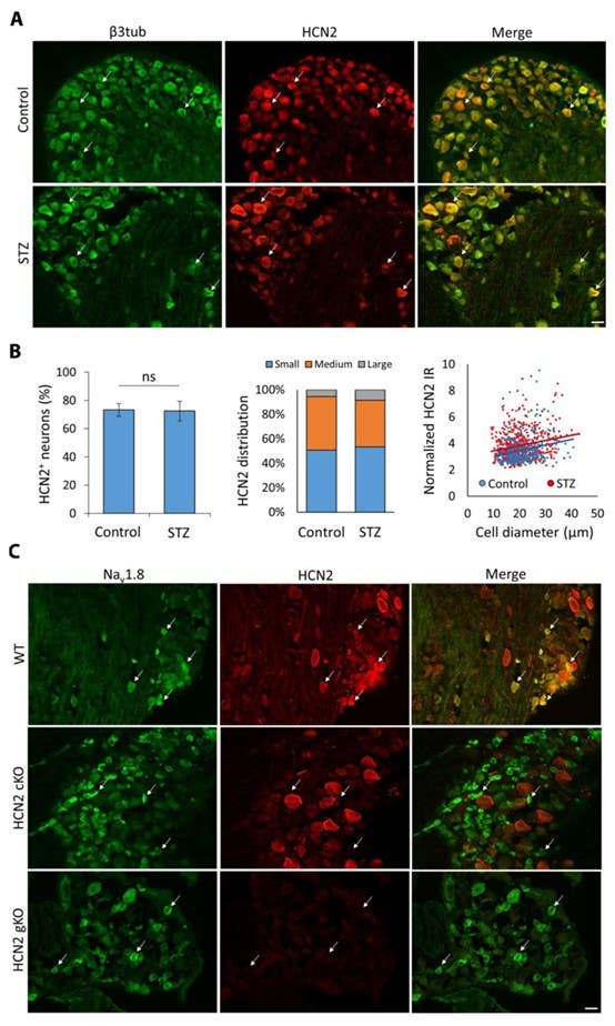
Figure 2. HCN2 expression in sensory neurons is not regulated by diabetes. (A) Representative immunohistochemical staining for the pan-neuronal marker β3-tubulin (β3tub, green), HCN2 (red), and the merged signal in transverse sections of lumbar DRG from control or diabetic (STZ) wild-type (WT) mice. Examples of HCN2+ neurons are indicated by arrows. Scale bar = 40 μm. (B) Left, percentage of HCN2+ neurons in control and STZ mice (Student’s t-test). Middle, distribution of HCN2+ neurons amongst DRG size classes (chi-square test). Right, intensity of HCN2 immunoreactivity (IR) as a function of neuronal size (linear regression analysis comparing slopes). Control (n=4 mice), STZ (n=4 mice). (C) Representative immunohistochemical staining in transverse sections of lumbar DRG for Nav1.8+ (marker of small nociceptive DRG neurons, green), HCN2 (red), and the merged signal in WT, HCN2 conditional knockout (cKO), and HCN2 global knockout (gKO). Examples of Nav1.8+ neurons are indicated by arrows. Scale bar = 40 μm.
Figure and legend adapted from Tsantoulas et al. (2017). DOI: 10.1126/scitranslmed.aam6072.
Using this validated approach, the authors found that neither the proportion of HCN2+ DRG neurons nor staining intensity differed between diabetic and control mice. The result shifted attention away from channel abundance and toward channel modulation. In diabetic neuropathy, HCN2 was not being upregulated; existing channels were being driven harder by elevated intracellular cAMP. The mechanism was not increased expression but functional potentiation of channels already present. Blocking HCN channels pharmacologically with ivabradine suppressed mechanical allodynia in both type 1 and type 2 diabetic animals. Genetic deletion of HCN2 specifically in small nociceptors produced the same effect and also reversed elevated C-FOS expression in dorsal horn neurons, indicating reduced nociceptive drive.
The mechanism chain was identical to the one Emery et al. had described: elevated cAMP, HCN2 potentiation, repetitive firing, pathological pain. What varied was the upstream source of the cAMP elevation. Three pain conditions—inflammatory hyperalgesia, neuropathic pain after nerve injury, and diabetic neuropathic pain—all required HCN2 in small nociceptors and all operated through the same cAMP-CNBD axis.
The Gap the Biology Created
This is where the story encounters a problem of target specificity. The genetic evidence for HCN2 as a pain driver is among the cleanest in peripheral pain biology. Relevant phenotypes can be abolished by isoform-specific deletion in defined cell types without disrupting normal acute sensation.
The pharmacology, however, has not kept pace with the genetics. The two HCN blockers most studied in pain models, ZD7288 and ivabradine, are not selective for HCN2. Ivabradine is used clinically for heart failure and stable angina precisely because it inhibits the HCN4-driven pacemaker current in sinoatrial node cells. Systemic HCN blockade that includes HCN4 is not a route to safe chronic analgesia.
Allosteric approaches targeting the CNBD interaction are therefore attracting increasing attention. In 2026, Loya-López and colleagues published direct proof of the cAMP-HCN2 causal link using TRIP8bnano, a synthetic peptide derived from the brain HCN auxiliary protein TRIP8b (4). TRIP8bnano selectively blocked cAMP access to the CNBD without altering baseline voltage sensitivity. Delivered as a plasmid to rat DRG neurons via intrathecal injection, it significantly reduced both mechanical allodynia and thermal hyperalgesia in a spinal nerve ligation model. The analgesic effect peaked at 24 hours and declined by 72 hours, consistent with the expression kinetics of the transfection system.
The authors acknowledged both the transient nature of the effect and the cardiac risks of approaches that might also reach HCN4. The concession is not that HCN2 is the wrong target, but that the target specificity achieved biologically—isoform-selective and cell-type-restricted—has no pharmacological equivalent yet.
What the Antibodies Were Measuring
Returning to the 2012 Acosta et al. study, it is worth considering what the antibodies were actually contributing to the science. What they provided was not evidence that HCN2 mattered, but evidence regarding where it mattered. They transformed HCN2 from a transcript detected in mixed DRG populations into a protein mapped to specific neuronal subtypes under defined pathological conditions.
HCN2 transcriptional data existed before then, but mRNA expression in mixed DRG populations cannot reveal which protein is present in which membrane, at what density, or how that distribution changes during disease. The immunostaining, validated through blocking peptides, isoform specificity controls, and HCN2 knockdowns, produced the protein distribution map that made the subsequent genetics interpretable. HCN2 increased in small nociceptors after inflammation while HCN1 did not. Without that distinction, the decision to build a nociceptor-specific HCN2 knockout rather than an HCN1 or pan-HCN deletion would have lacked its anatomical rationale.
Reagents play only a small part in scientific reasoning, but the measurements they enable propagate into hypothesis-level decisions. The antibody characterization in 2012 may not have been the experiment that changed the field, but it was the measurement that made the field-changing experiments possible.
The contribution of HCN2 immunostaining extended beyond the original localization findings. In the Acosta et al. study, subtype-specific antibodies revealed a transient increase in HCN2 expression in small nociceptors after inflammation. In the diabetic neuropathy study by Tsantoulas and colleagues, the same anti-HCN2 antibody was validated using both nociceptor-specific (NaV1.8-HCN2−/−) and HCN2 gKO mice before being used to compare diabetic and control tissue. HCN2 immunoreactivity was selectively lost from small nociceptors in the HCN2 cKOs and absent altogether in the HCN2 gKOs, providing strong genetic confirmation of staining specificity. Rather than detecting increased expression in diabetic animals, the study showed that HCN2 protein levels remained unchanged despite robust pain behavior.
Together, these findings helped distinguish two routes to HCN2-dependent pain: one involving altered protein abundance after inflammation and another involving functional potentiation of existing channels through elevated cAMP. In both cases, protein-level measurements helped define the mechanism.
Coming Back to Ih
For years, the Ih in nociceptors was treated as a minor electrophysiological feature—present, measurable, but not especially important. The work that followed showed otherwise. Across inflammatory, neuropathic, and diabetic pain models, a consistent picture emerged: pathological pain depends not on the Ih in general, but on HCN2 operating within a specific cellular and signaling context.
That insight did not come from a single experiment. It emerged through the combination of protein localization, conditional genetics, and mechanistic physiology. Immunostaining identified where HCN2 was expressed and how that expression changed; genetics established causality; functional studies revealed the cAMP-dependent mechanism linking HCN2 to persistent pain.
The biology is now unusually clear. The remaining challenge is pharmacological: developing therapies capable of matching the precision that the underlying science has already achieved. If that challenge can be met, the channel once dismissed as background noise may become one of the most compelling targets in peripheral pain medicine.