Interrogation of cleared organs often produce misleading results – here’s how to correct it.
There’s a moment in every 3D imaging experiment where you pull up the confocal stack and it looks, genuinely, spectacular. A whole cleared kidney rotating on your screen with the arterial bed threading through it like copper wire. You’ve spent days or weeks on this.
Then you realize a large portion of what you’re looking at is autofluorescence. Just the tissue itself, radiating green noise at 500–550 nm because that’s what biological tissue does – metabolic byproducts, structural proteins, lipofuscin, flavins, all doing their level best to ruin your day.
While we’ve all been in this situation, this is a problem that’s been lurking in the 3D imaging field for years. However, most researchers have come to terms with it or figured out how to just workaround it. We lower laser power, choose our fluorophores carefully, and just accept that our organ images are going to look like they’re lit from inside. That’s the standard approach and it’s not acceptable.
The Thing You’ve Been Accepting
Autofluorescence does something specific and annoying to 3D imaging data that goes beyond aesthetics. It drags down your SBRs, which means it’s actively hiding biology. Antigens expressed at low levels become invisible against the noise. Moreover, multiplex imaging, which requires distinguishing actual signals across multiple channels, becomes an exercise in frustration when the background is competing with the signal.
Human tissue makes all of this worse. Human organs are more fibrous, more pigmented, more autofluorescent, and generally less cooperative than murine equivalents. Anyone who’s tried to optically clear a human biopsy specimen knows this all too well. The clearing protocols that work beautifully on a mouse heart will do about half the job on human tissue, and the autofluorescence that was merely annoying in the mouse model becomes genuinely limiting in the human specimen. So, the problem is real, widespread, and it’s been limiting what 3D imaging can actually tell us.
What Someone Actually Did About Autofluorescence
One group decided they’d had enough of just coming to terms with autofluorescence. A paper published in Cell Reports Methods (1) describes a technique referred to as hydrogen peroxide (H2O2)-sodium azide (NaN3)-DMSO (HyPer-3D), and which turns out to be a surprisingly simple solution to a problem that’s been frustrating scientists for years.
The logic is elegant. H2O2 has long been used for tissue decolorization and autofluorescence quenching. The problem is that tissue contains catalase, which converts H2O2 into water and oxygen, and the resulting oxygenation causes tissue damage, and damaged tissue regions then exhibit increased autofluorescence.
The fix, it turns out, is NaN3, which inhibits catalase activity, moderating oxygen production without halting it entirely. This lets the decolorization proceed without the tissue tearing. DMSO acts as a permeabilizing agent, enabling the treatment to penetrate thicker sections and whole organs. The whole thing runs in a basic buffered solution at pH 9.5. That’s it – simple.
But what this achieves, according to the paper, is substantial:
- SBRs increased by up to 30-fold in whole murine organs using light sheet microscopy.
- Optical clearing capacity potentiated by up to 6-fold across all nine clearing protocols tested including: EZ Clear, CUBIC, iDISCO+, ScaleS, PACT, SHANEL, MACS, SeeDB, and glycerol.
- Detection of poorly recognized antigens improved by up to 4 to 5-fold.
- Fluorescent reporter detection increased by up to 5-fold.
That last point is especially relevant. Fluorescent reporter proteins are routinely diminished by optical clearing protocols – the assumption has become that you’ll lose some of your GFP, Venus, or tdTomato signal in the clearing process, and you make your peace with it. HyPer-3D actually improves reporter detection, because most of the signal was masked by background noise.
What HyPer-3D Enabled
The SBR improvements translate directly into biology you couldn’t see before. The paper demonstrates 3D imaging of previously undocumented nephron segments – the distal convoluted and connecting tubules, which are the most autofluorescent structures in an organ that is itself notorious for autofluorescence. With HyPer-3D, these segments become resolvable at single-cell resolution. The imaging also enabled AI-based autosegmentation of renal tubules, the sort of approach that requires clean signal to work, and will simply fail if the data are too noisy.
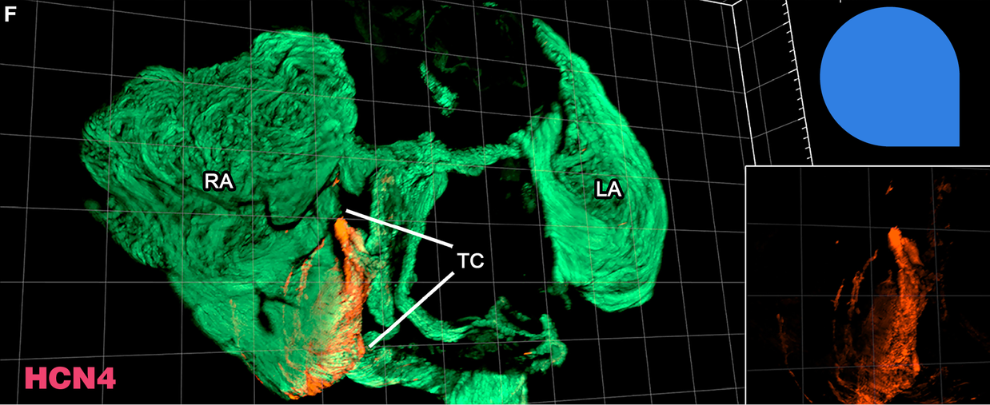
In cardiac tissue, HyPer-3D allowed for the visualization of the sinoatrial node (SAN), a small cluster of specialized pacemaker cells located in the right atrium, near the opening of the superior vena cava. It acts as the heart’s natural pacemaker, generating electrical impulses that initiate each heartbeat and set the overall heart rate. Visualizing the sinoatrial node is inherently challenging because it is not a distinct, well-defined structure but rather a small, diffuse collection of specialized cells embedded within the atrial wall. Its exact location varies between individuals, and it lacks clear anatomical boundaries or visible landmarks that would make it easy to pinpoint during imaging. To visualize the SAN, the researchers used Alomone’s Anti-HCN4 Antibody (#APC-052), and it performed well (Figure 1). However, the more interesting finding was that HyPer-3D produced a clearly defined HCN4+ node at the terminal crest – the correct anatomical site – that standard H2O2 treatment, the existing gold standard, failed to reveal with comparable clarity. HyPer-3D was successful at achieving a clear signal, even though whole-tissue imaging of the SAN is difficult because the pacemaker signal tends to be diffuse and easily lost against cardiac autofluorescence.
In testis tissue, the use of HyPer-3D to detect spermatogonial stem cell markers (cKIT, STRA8, and GFRα1) – markers that are poorly expressed and easily lost against background – became distinguishable with 3 to 4.5-fold higher SBRs. The paper then showed 3D imaging of A-paired and A-aligned spermatogonia with enough resolution to count cells in chains. This is the kind of imaging that was theoretically possible before but practically very difficult to obtain until HyPer-3D was developed.
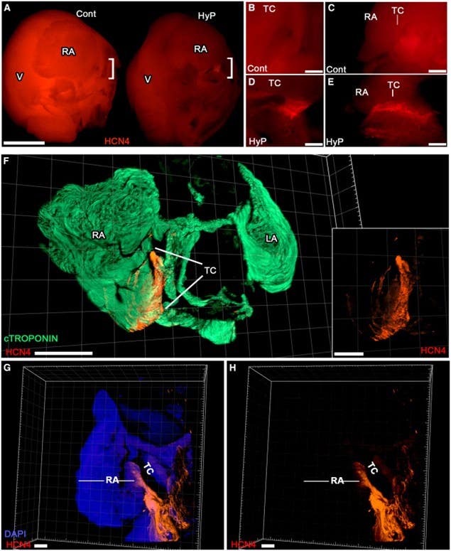
Figure 1. 3D interrogation of muscular organs with enhanced clarity. (A–E) Immunofluorescence performed on whole atria for HCN4 (red) that marks the sinoatrial node (SAN). Atria processed using standard hydrogen peroxide (H2O2) treatment (control, Cont) or HyPer-3D (HyP) were imaged side-by-side for comparison (A, scale bars represent 2 mm; RA, right atria; LA, left atria; V, ventricle. High-magnification analyses of bracketed regions in (A) showed a distinct HCN4+ node at the terminal crest (TC) of hearts imaged with HyPer-3D (D, coronal view; E, lateral view), which was not readily discernible in control hearts (B, coronal view; C, lateral view). Scale bars in B-E represent 0.25 mm. (F) 3D confocal microscopy imaging of formamide/glycerol-cleared whole atria immunolabeled for both the working myocardium cTROPONIN-T ( green), and HCN4 pacemaker channels (red; scale bars, 1 mm). The inset shows SAN alone. (G and H) Staining with DAPI and HCN4 (G, merged); H, HCN4 alone; scale bars represent 0.2 mm).
Image taken from Choi et al. (2026). https://doi.org/10.1016/j.crmeth.2026.101375.
The Human Tissue Problem, Partially Solved
For human specimens, the authors developed HyPer-3D + Blue light (BL): the standard treatment combined with blue light exposure (470/40 nm excitation). This method was designed to address persistent green channel autofluorescence, which proved more resistant to chemical quenching alone. Green autofluorescence in human kidney tissue was abolished in 4 hours. The combination yielded SBRs that were 5-fold higher than untreated controls in the green channel when used with the SHANEL clearing protocol and allowed multiplex imaging of human glomerular architecture with cellular resolution.
The honest caveat from the paper itself: the mechanism by which green and red autofluorescence differ in nature remains an open question. The method works empirically, but the mechanistic explanation for the difference between green and red autofluorescence is for future research.
So What Do You Do With This?
The straightforward answer is that HyPer-3D is a pre-treatment step that may be incorporated into your existing clearing workflow before you do anything else. You don’t need to switch protocols or acquire new equipment. The reagents – H2O2, NaN3, DMSO, Triton X-100, Tris-HCl, and potassium chloride – are standard shelf items. The starting concentration is 0.05% H2O2, titrated upward if needed, with a maximum of 0.5% across all tissues tested. Simply monitor for oxygenation and if it gets excessive, cold PBS stops it.
The one thing requiring attention is familiarity with your tissue type. Different organs, different species, different fixation histories, all affect how the reaction proceeds. The paper is honest about this as they explain user familiarity as a genuine variable, especially early on. The full protocol is published in the STAR Methods section of the paper so you can easily follow it if you want to try HyPer-3D.
The Uncomfortable Part
There is something slightly awkward about all of this. A 30-fold improvement in SBRs, all from a combination of three reagents in a basic buffer. The biological structures this reveals were always there – the pacemaker cells, the spermatogonial stem cell chains, the connecting tubule architecture – they were present in every tissue block that went through clearing over the past decade without HyPer-3D. We just couldn’t see them properly.
That’s not a criticism of the field. Optical clearing is a young methodology and it’s been advancing fast, but it does suggest that a meaningful fraction of 3D imaging data in the literature was collected with a significant constraint on what could be detected. And that the bar for “acceptable background” was set by what the tools could achieve, not by what the biology required.
HyPer-3D raises that bar and whether the field will uniformly adopt a pre-treatment step that adds time and requires some practice is a different question. That’s not going to depend on the data alone.


 Antibody")
 Antibody")