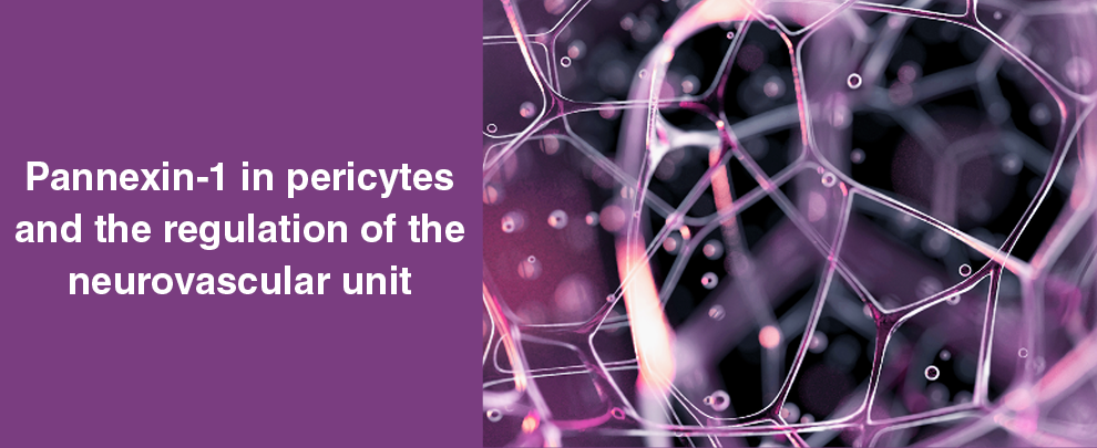
The brain must continuously adjust blood flow to match neuronal activity. Most work on this problem has focused on arterioles and smooth muscle. But capillaries account for much of cerebrovascular resistance, and pericytes – contractile cells positioned along capillary walls – actively control capillary diameter. What has been missing is a molecular mechanism that explains how these cells translate local biochemical signals into changes in vascular tone.
A recent study from Universidad de la República, Uruguay, showed that pannexin-1 (Panx1) channels expressed in pericytes acted as a local regulator of capillary diameter, linking ATP signaling, neuronal activity, and cerebral blood flow (1).
The work revealed a signaling circuit in which Panx1-mediated ATP release controled pericyte calcium signaling and contractility. Disrupting that circuit altered capillary dynamics and impaired memory performance in mice.
Pericytes Express Functional Panx1 Channels
The starting point of the study was focused on a basic question: do pericytes express active Panx1 channels? Using acute hippocampal slices, the authors measured uptake of the membrane-impermeant dye, ethidium bromide (Etd⁺), a common indicator of large-pore channel activity. Pericytes showed basal dye uptake under resting conditions, indicating constitutively open membrane pores. Pharmacological inhibition of Panx1 reduced uptake substantially, while connexin-specific inhibitors had little effect.
Genetic models confirmed the results since both global Panx1 knockout mice and mice with pericyte-specific Panx1 deletion showed markedly reduced dye uptake in pericytes.
Immunofluorescence experiments demonstrated Panx1 localization in pericytes within the hippocampus. Detection of the channel relied on our Anti-Pannexin 1 Antibody (#ACC-234), with knockout tissue serving as a control for specificity (Figure 1). These experiments confirmed that Panx1 protein is present in pericytes alongside its expression in endothelial cells, smooth muscle cells, and neurons within the neurovascular unit.
Taken together, pharmacological, genetic, and immunological approaches converge on the same conclusion: brain capillary pericytes express active Panx1 channels under resting physiological conditions.
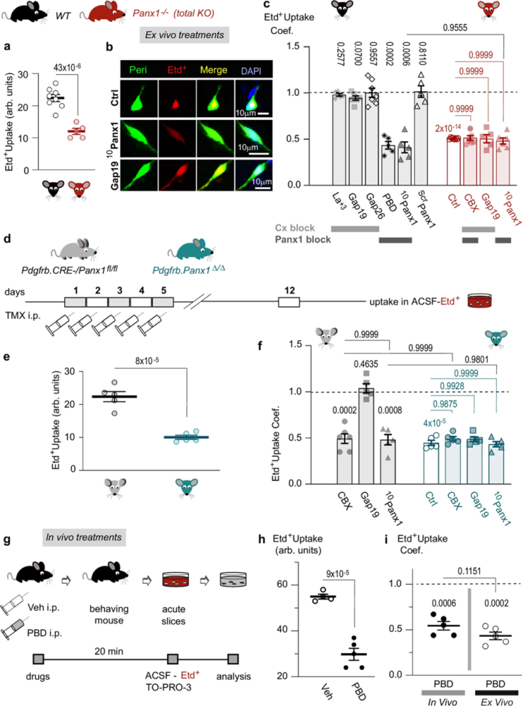
Figure 1. Panexin 1 channels expressed by hippocampal pericytes are functional in resting conditions and mediate a molecular exchange with the cerebral microenvironment. a Ethidium bromide (Etd+) uptake [arbitrary units(arb.)] by hippocampal pericytes from global knockout (KO) Panx1−/− mice (red; n = 5) is decreased relative to wild-type (WT) mice (black; n = 8); unpaired two-tailed t-test. b Fluorescent microphotographs show the mimetic peptide 10Panx1, which inhibits pericyte Etd+ uptake in WT hippocampal slices. c Etd+ uptake coefficients (Coef.) of pericytes from WT hippocampal slices are insensitive to Cx blockers, La3+ (n = 5), Gap19 (n = 6), Gap26 (n = 7), and scrambled peptide ScrPanx1 (n = 5). However, the Etd+ uptake does respond to Panx1 blockers PBD (n = 5) and 10Panx1 (n = 5) relative to the Ctrl (dashed line; n = 8). The pericytes from Panx1−/− KO mice are unresponsive to all blockers, including CBX (n = 8), Gap19 (n = 5) and 10Panx1 (n = 6) compared to the Ctrls (n = 8). All data were normalized relative to control WT mice. d Tamoxifen (TMX) administration i.p. to Pdgfrb/Panx1Δ/Δ and Pdgfrb.CRE-/Panx1fl/fl littermate mice. e Etd+ uptake (arb. units) by pericytes in hippocampal slices from conditional Pdgfrb/Panx1Δ/Δ mice (n = 5) is decreased relative to Pdgfrb.CRE-/Panx1fl/fl littermates (n = 5); unpaired two-tailed t-test. f The Etd+ uptake Coef. of pericytes from Pdgfrb.CRE-/Panx1fl/fl slices is inhibited by CBX (n = 6) and 10Panx1 (n = 5) compared to control (dashed line; n = 6) but remains unresponsive to Gap19 (n = 5). In contrast, Etd+ uptake by hippocampal pericytes from Pdgfrb/Panx1Δ/Δ mice is insensitive to all blockers, including CBX (n = 5), Gap19 (n = 5), and 10Panx1 (n = 5), compared to the respective Ctrls (n = 5). All data were normalized relative to the Ctrl Pdgfrb.CRE-/Panx1fl/fl mice. g I.p. administration of vehicle (Veh) 10 ml/kg and PBD 200 mg/kg to WT mice. h Etd+ uptake (arb. units) by hippocampal pericytes from PBD-treated mice (n = 5) decreased compared to Veh-treated mice (n = 4); unpaired two-tailed t-test. i Etd+ uptake Coefs. of hippocampal pericytes after in vivo treatments [Veh (dashed line), n = 4; PBD, n = 5] and ex vivo treatments [Ctrl (dashed line), n = 7; PBD, n = 5], showed similar effects. Two-tailed one-sample t-tests were used to compare PBD to Veh (in vivo) or Ctrl (ex vivo) (over bars). In a, c, e, f, h, and i, the data are presented as the mean ± SEM. n represents the number of mice.
Image taken from Mai-Morente et al. (2025). https://doi.org/10.1038/s41467-025-61312-0.
Panx1 Drives ATP Signaling in Pericytes
Panx1 channels are known to release ATP in many cell types. The authors therefore asked whether pericyte Panx1 contributes to local purinergic signaling.
In cultured cerebral pericytes, inhibiting Panx1 markedly reduced extracellular ATP levels. The same manipulation lowered baseline intracellular Ca²⁺ signals, suggesting that ATP released through Panx1 channels contributes to tonic purinergic signaling within these cells.
The data supported a feedback mechanism: ATP activated purinergic receptors, which in turn stimulated additional Panx1 channel activity and ATP release. This amplified Ca²⁺ signaling within pericytes and promoted their contractility.
Purinergic Receptors Regulate Panx1 Activity
To identify the receptors involved, the study examined purinergic signaling upstream of Panx1. Pharmacological inhibition experiments showed that P2X7 and P2Y6 receptors regulate Panx1 activity in pericytes. Blocking either receptor reduced Panx1-dependent dye uptake, and combined inhibition produced an even stronger effect.
Immunofluorescence staining revealed that both receptors were localized on pericyte membranes in hippocampal tissue. These experiments used Alomone’s rabbit polyclonal antibodies [Anti-P2X7 Receptor Antibody (#APR-004) and Anti-P2Y6 Receptor Antibody (#APR-011)] to confirm receptor localization within the neurovascular unit.
The emerging mechanism was revealed as a purinergic signaling loop: ATP activated P2X7 and P2Y6 receptors, receptor signaling opened Panx1 channels, and Panx1 released more ATP. This positive feedback increased intracellular Ca²⁺ levels and strengthened pericyte contraction.
Pericyte Panx1 Controls Capillary Diameter
If Panx1 regulates ATP levels and Ca²⁺ signaling in pericytes, it should also influence capillary tone.
The authors measured capillary diameter in hippocampal slices relative to the position of pericyte somata. Manipulating extracellular ATP levels produced predictable vascular responses:
- Removing ATP with apyrase caused capillary dilation near pericyte cell bodies
- Adding ATP induced capillary constriction, again strongest near pericytes
Both responses depended on Panx1 expression in pericytes. When Panx1 was genetically deleted in these cells, ATP-driven changes in capillary diameter were strongly reduced.
These findings suggested that Panx1 amplified local ATP concentrations near pericytes, enabling purinergic signaling to effectively control capillary resistance.
Neuronal Activity Suppresses Panx1 to Dilate Capillaries
The study then examined how neuronal signaling interacts with this pathway. Glutamate – the brain’s principal excitatory neurotransmitter – reduced Panx1 channel activity in hippocampal slices, an effect partially rescued by NMDA and AMPA receptor antagonists. However, cultured pericytes were unresponsive to glutamate, which indicated the signal is unlikely to act directly on pericyte Panx1. The authors proposed that NMDA/AMPA activation triggers the release of diffusible messengers – i.e., nitric oxide and arachidonic acid – from neurons and glia, and that these intermediaries inhibit pericyte Panx1 function.
The physiological consequences followed the expected outcome. Glutamate exposure increased capillary diameter by 18% near pericytes, consistent with functional hyperemia during neuronal activity.
Deleting Panx1 in pericytes abolished this glutamate-evoked dilation, indicating that Panx1 acted as the vascular control point through which neuronal signals modulate capillary tone.
Panx1 Activity Influences Memory Performance
The study extended these findings to behavior analysis. Mice lacking Panx1 specifically in pericytes showed impaired performance in two cognitive tasks: the object location test, which depends on hippocampal function, and the novel object recognition test, which engages cortical circuits. Both assays measure memory-dependent exploration behavior, and performance was reduced in Panx1-deficient mice relative to controls.
The authors proposed that during learning, pericyte Panx1 supports cognition by enabling activity-dependent capillary dilation and local increases in blood flow.
A Molecular Control Point in the Neurovascular Unit
Taken together, the findings place pericyte Panx1 at a crucial point in neurovascular regulation. Panx1 channels release ATP from pericytes, which activates purinergic receptors and amplifies Ca²⁺ signaling to regulate contractility. Neuronal glutamatergic signaling suppresses this pathway, allowing capillary dilation during periods of neural activity.
Through this mechanism, a membrane channel in pericytes links neuronal signaling, capillary blood flow, and behavioral outcomes in the brain.