One Marker Doesn’t Cut It
Single-marker antibody labeling might seem convenient, but it rarely provides a clear read on microglia. These resident CNS immune cells share markers with other myeloid populations and shift their phenotype depending on age, region, or disease. The result of relying on a single marker? False positives, misclassification, and lost resolution. Instead, combining antibodies against multiple markers will give you the specificity needed for accurate microglial detection.
Where Single Markers Fall Short
Ionized calcium-binding adaptor molecule 1 (Iba1) remains a common marker for microglia detection. But it’s not exclusive. Border-associated macrophages (BAMs) and infiltrating monocytes also express Iba1 (1), and under inflammatory conditions, these cells can enter the parenchyma and adopt microglia-like morphology (2). Thus, while Iba1 stains plenty of cells, it doesn’t tell you which ones are truly microglia.
CD45, a membrane-bound tyrosine phosphatase, is commonly used to distinguish microglia (CD45lo) from other immune cells (CD45hi). That works – until it doesn’t. In disease or injury, microglial CD45 expression rises (3), which narrows the gap and makes it harder to distinguish populations by intensity alone.
And even if your marker were microglia-specific, there’s still the issue of timing and context. Marker expression varies across brain regions and developmental windows (4). A marker that labels microglia in the adult cortex might miss them in the neonatal brainstem, or in an inflamed hippocampus.
Transmembrane Protein 119 (TMEM119) Offers Better Specificity – Within Limits
Bennett et al. identified TMEM119 as a reliable microglia-specific marker that is expressed in both mouse and human microglia. TMEM119 is absent from BAMs and infiltrating monocytes, even during inflammation (4). Antibodies directed against TMEM119 allow accurate labeling in fixed sections, and when conjugated to fluorophores, support live-cell detection in flow cytometry or imaging.
But even TMEM119 isn’t a universal solution. Its expression is developmentally regulated and becomes stable only by the second postnatal week in mice (4). It can also be downregulated under certain disease conditions, reducing its reliability in those settings.
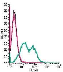
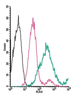
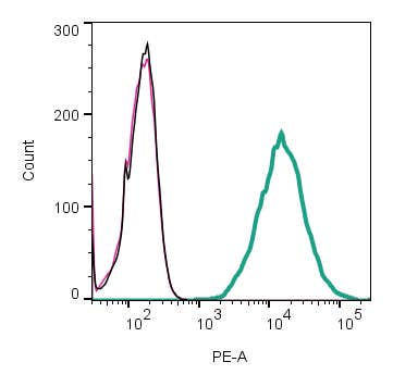
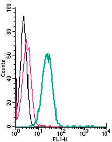
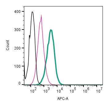
We offer an anti-TMEM119 antibody that specifically binds to the extracellular region of the protein (Figure 1). This allows for live-cell labeling and flow cytometry, as well as standard immunohistochemistry, immunocytochemistry, and Western blotting. To simplify live-cell labeling, we supply the anti-TMEM119 antibody conjugated to a range of fluorophores, eliminating the need for secondary antibodies. We also offer unconjugated TMEM119 antibodies. The following anti-TMEM119 antibodies are available from Alomone Labs:
- Anti-TMEM119 (extracellular) Antibody (#ANR-175), unconjugated
- Anti-TMEM119 (extracellular)-APC Antibody (#ANR-175-APC), conjugated to allophycocyanin (APC)
- Anti-TMEM119 (extracellular)-PE Antibody (#ANR-175-PE), conjugated to R-phycoerythrin (PE)
- Anti-TMEM119 (extracellular)-FITC Antibody (#ANR-175-F), conjugated to fluorescein isothiocyanate (FITC)
Combining Antibodies Gives You Confidence
Specificity improves when antibodies are used in combination. A common flow cytometry strategy, for example, uses CD11b and CD45 to gate myeloid cells, then adds TMEM119 to pinpoint microglia. In this setup, microglia are CD11b+ CD45lo TMEM119+, while monocyte-derived cells are CD45hi TMEM119− (5).
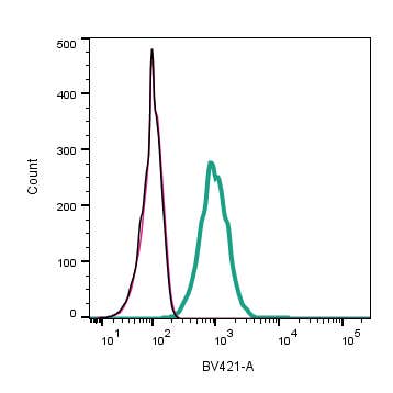
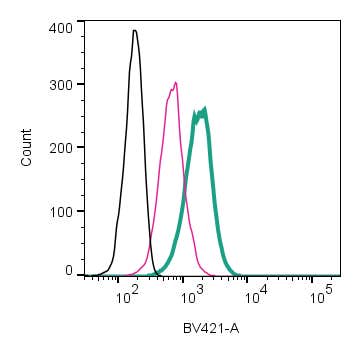
Markers such as P2Y12 receptor and Sall1, microglial transcription factor, can further refine this distinction, depending on the system (5, 6). Alternatively, you can combine P2Y12 receptor with TMEM119 to identify discrete cell populations (shown in Figure 1) using our Anti-P2Y12 Receptor (extracellular)-mFluor™ Violet 450 Antibody (#APR-020-V), which is directly conjugated to mFluor™ Violet 450, and our Anti-TMEM119 (extracellular)-PE Antibody (#ANR-175-PE), which is directly conjugated to PE fluorophore.
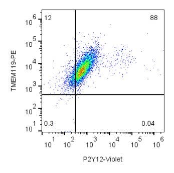
Figure 1. Live intact mouse BV-2 microglia cells were simultaneously stained with Anti-P2Y12 Receptor (extracellular)-mFluor™ Violet 450 Antibody (#APR-020-V) and Anti-TMEM119 (extracellular)-PE Antibody (#ANR-175-PE) followed by analysis in a flow cytometer.
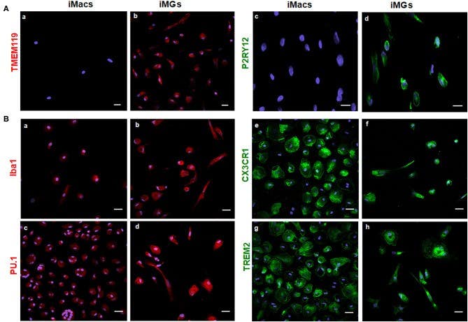
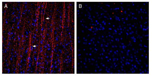
Figure 2. Induced microglia (iMG) cells express microglia-specific markers (TMEM119 and P2RY12) and other myeloid-specific markers. Immunocytochemistry shows induced macrophages (iMacs) and iMGs derived from peripheral monocytes. A) iMG cells express unique microglial markers TMEM119 (b) and P2RY12 (d) stained with Alomone Labs’ Anti-P2Y12 Receptor (extracellular) Antibody (#APR-020). These markers were absent from iMacs (a,c). B) Several myeloid lineage markers were expressed on both iMacs and iMG cells, such as Iba1 (a,b), PU.1 (c,d), CX3CR1 (e,f), and TREM2 (g,h). Image adapted from Banerjee, et al. 2021 (7).
We offer the following extracellular anti-P2RY12 antibodies in multiple directly conjugated forms and unconjugated forms:
- Anti-P2Y12 Receptor (extracellular) Antibody (#APR-020)
- Guinea Pig Anti-P2Y12 Receptor (extracellular) Antibody (#APR-020-GP)
- Anti-P2Y12 Receptor-ATTO Fluor-594 Antibody (#APR-012-AR), conjugated to ATTO-594 fluorescent dye
- Anti-P2Y12 Receptor (extracellular)-FITC Antibody (#APR-020-F)
- Anti-P2Y12 Receptor (extracellular)-mFluor™ Violet 450 Antibody (#APR-020-V)
- Anti-P2Y12 Receptor (extracellular)-PE Antibody (#APR-020-PE)
- Anti-P2Y12 Receptor (extracellular)-APC Antibody (#APR-020-APC)
Fluorophore-conjugated antibodies allow simultaneous detection of these markers in multiplex panels, making them useful not only for live sorting, but also for resolving subtle shifts in microglial state. A well-designed panel will allow you to:
- Distinguish microglia from morphologically similar myeloid cells
- Track microglial maturation and activation over time
- Isolate pure microglia populations for transcriptomic or functional studies
Your choice of antibody panel should match the context: developmental stage, species, brain region, and disease model.
Why This Matters for Experimental Design
When it comes to correctly identifying cells like microglia, multiple antibodies should be your baseline approach. Without a panel, you risk misidentifying cells – especially in neuroinflammatory models, where infiltrating cells and activated microglia overlap in both morphology and marker profile.
Well chosen antibody combinations offer clear advantages. They help define microglial populations accurately. They let you track state changes over time. They also ensure high-confidence isolation of live microglia for downstream applications such as RNA-seq or functional assays.
The right antibodies – used together, not alone – make the difference between guessing and knowing.
The Power of Microglial Paneling
The ability to combine multiple microglial markers in a well-structured antibody panel opens the door to deeper, more precise analyses. Beyond simple identification, panel-based approaches enable dynamic profiling of microglial activation states, region-specific differences, and functional transitions. By pairing carefully selected, directly conjugated antibodies, researchers can achieve cleaner data, stronger confidence, and a more complete understanding of microglial biology.
Resources
You can find our full collection of antibodies directed against microglia markers – both conjugated and unconjugated – here.
If you need to compare fluorophore spectra or check for potential overlap, our Spectra Viewer gives you everything in one place. Plot excitation and emission curves, adjust filters, and see where things might clash. It’s a clear, visual way to plan smarter panels and avoid compensation issues later on.
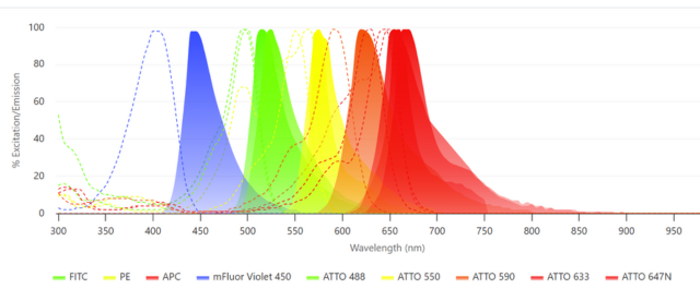
If you want to select antibodies that are conjugated to fluorophores, check out our Panel Builder. This will help you pick out the perfect antibody combination for assays such as flow cytometry or multiplex options. Simply choose your targets, pick your antibodies, and it maps out the best possible fluorophore combinations for your setup. Less guesswork, cleaner data.
Custom conjugation services are also available so you can get exactly what you need for your experiments.